What Knee Pain Biologic Therapy Can and Cannot Do
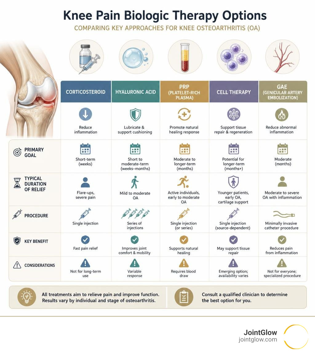
Knee pain biologic therapy includes injections and minimally invasive treatments designed to reduce inflammation, improve symptoms, or support joint function without knee replacement. The best-supported options today are corticosteroid injections for short-term relief, hyaluronic acid for some people with mild to moderate osteoarthritis, and platelet-rich plasma (PRP) for selected patients seeking longer-lasting symptom improvement.
These treatments may help you move with less pain, return to physical therapy, and sometimes postpone surgery. They do not reliably regrow a severely worn, bone-on-bone knee or guarantee that a future knee replacement will not be needed.
Knee osteoarthritis develops as cartilage loses its ability to cushion the joint. Inflammation, swelling, changes in the joint lining, and altered pain nerves can make every step harder. It is increasingly common: knee osteoarthritis prevalence rose about 48% from 1990 to 2019.
Before choosing a biologic option, ask a qualified clinician four practical questions:
- What is causing my pain: arthritis, inflammation, a meniscus problem, poor alignment, or something else?
- How advanced is my osteoarthritis on X-ray or MRI?
- Which treatment has evidence for someone with my age, weight, health conditions, and activity goals?
- Is it approved, safely prepared, and covered by my insurance?
This guide explains the science, evidence, recovery, cost questions, and realistic expectations behind biologic knee injections and other emerging non-surgical therapies.
Understanding Knee Pain Biologic Therapy and Core Mechanisms
Knee osteoarthritis (OA) affects an estimated 5 million individuals in the United States alone. Projections indicate that total knee arthroplasty (TKA) procedures will increase by 69% by 2050 compared to 2012 levels. This substantial demand has accelerated clinical interest in orthobiologic therapies designed to preserve joint integrity, modulate biochemical pathways, and manage debilitating discomfort.
Orthobiologics are naturally derived biological substances—including blood fractions, concentrated cellular products, tissue grafts, and protein hydrogels—administered directly into the intra-articular space. Unlike traditional analgesics that temporarily blunt systemic pain pathways, biological interventions aim to alter the intra-articular microenvironment. Degenerative joint disease is not merely mechanical “wear and tear”; it is an active, cytokine-driven inflammatory cascade. Interleukin-1 beta (IL-1β), tumor necrosis factor-alpha (TNF-α), and matrix metalloproteinases (MMPs) actively degrade extracellular type II collagen and aggrecan (Canada et al., 2019).
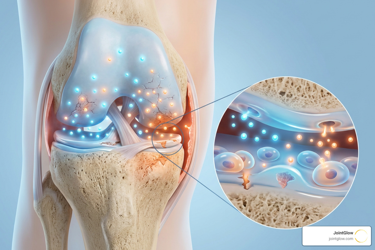
Biologic therapies deliver concentrated growth factors, anti-inflammatory cytokines, and bioactive scaffolding directly to the damaged synovium and articular cartilage. As highlighted in literature assessing whether orthobiologics represent a dream or reality for knee osteoarthritis, their therapeutic effect relies primarily on paracrine signaling. Rather than spontaneously turning into pristine new cartilage, these injected biological agents stimulate native chondrocytes, inhibit destructive proteases, downregulate inflammatory mediators, and promote vascular homeostasis within the joint capsule.
Platelet-Rich Plasma (PRP) Injections
Platelet-Rich Plasma (PRP) is an autologous biologic therapy prepared by drawing a patient’s peripheral blood and centrifuging it to concentrate platelets 3 to 5 times above baseline physiological levels. These concentrated platelets contain alpha-granules loaded with potent signaling proteins, including Transforming Growth Factor-Beta (TGF-β), Platelet-Derived Growth Factor (PDGF), Vascular Endothelial Growth Factor (VEGF), and Fibroblast Growth Factor (FGF).
Upon intra-articular injection, these granules degranulate, releasing biomolecules that suppress nuclear factor kappa B (NF-κB) activation in synovial cells. This action interrupts the cycle of synovial inflammation and cartilage breakdown. Comparative studies show that intra-articular platelet-rich plasma demonstrates superior clinical and biomarker outcomes compared with standard corticosteroids and non-steroidal anti-inflammatory drugs (NSAIDs) in late-stage knee osteoarthritis. In clinical trials, serial PRP injections have significantly lowered cartilage breakdown markers such as cartilage oligomeric matrix protein (COMP) and matrix metalloproteinase-3 (MMP-3), while increasing neuroprotective and anti-inflammatory markers like sTREM2 and TGF-β1.
For early to moderate OA (Kellgren-Lawrence grades I–II), PRP regularly outperforms viscosupplementation (hyaluronic acid) at 6- and 12-month follow-ups. Even in advanced structural disease (Kellgren-Lawrence grades III–IV), research indicates PRP can postpone the need for total knee arthroplasty for more than 1.5 years in 74.1% of patients, with a median surgical delay of 5.3 years.
Mesenchymal Stem Cell (MSC) and Cellular Therapies
Cellular therapies represent an advanced branch of regenerative orthopedics. They utilize multipotent progenitor cells harvested from various anatomical sources:
- Bone Marrow Aspirate Concentrate (BMAC): Harvested autologously from the iliac crest and centrifuged to isolate nucleated cells, platelets, and mesenchymal stem cells. Although MSCs make up only 0.001% to 0.01% of total cells in BMAC, the concentrate is rich in Interleukin-1 receptor antagonist (IL-1Ra), which blocks destructive IL-1 signaling.
- Adipose Stromal Vascular Fraction (SVF): Isolated from autologous lipoaspirate via enzymatic digestion or mechanical processing, yielding a high volume of pericytes, pre-adipocytes, and stromal cells.
- Umbilical Cord-Derived Mesenchymal Stem Cells (UC-MSCs): An allogeneic, “off-the-shelf” option derived from discarded umbilical cord tissue (Wharton’s Jelly). UC-MSCs possess high proliferative potential and low immunogenicity, bypassing the donor-site morbidity associated with bone marrow or fat harvesting.
The clinical efficacy of these therapies hinges on paracrine immunomodulation. Recent trials investigating multiple injections of umbilical cord mesenchymal stem cells for severe knee osteoarthritis demonstrated significant clinical gains (Tong et al., 2025). In a triple-blind trial, UC-MSC therapy reduced visual analog scale (VAS) pain scores from 7.2 to 3.36 at 12 months (a 3.84-point drop), compared to a decrease from 7.0 to 4.6 in the hyaluronic acid control arm. Furthermore, Western Ontario and McMaster Universities Osteoarthritis Index (WOMAC) scores decreased by 23.2 points in the UC-MSC cohort versus 15.6 points in controls, alongside structural improvements in magnetic resonance observation of cartilage repair tissue (MOCART) scores.
However, cellular orthobiologics show variability across clinical platforms. Large-scale multicenter trials analyzing cell-based versus corticosteroid injections have revealed that while BMAC, SVF, and UC-MSCs provide sustained symptom relief, they do not always achieve statistical superiority over traditional injections across broader, unstratified patient groups (Mautner et al., 2023). This highlights the importance of careful patient selection.
Amniotic and Collagen Hydrogel Injections
Amniotic suspensions derived from human placenta contain rich pools of growth factors (EGF, basic FGF, PDGF), structural proteins, and natural cytokine antagonists (IL-1Ra, IL-10). A double-blind, prospective trial showed that amniotic tissue injections are an effective alternative to corticosteroids, providing sustained improvements in Knee Injury and Osteoarthritis Outcome Scores (KOOS) and functional capacity over 12 months, avoiding the tissue degradation risk associated with repeated steroid injections.
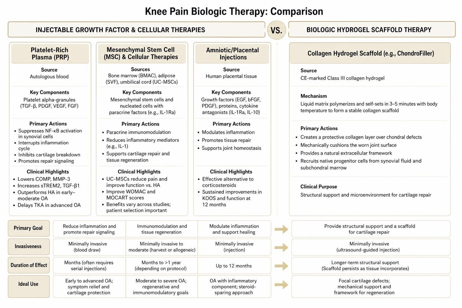
In parallel, structural biomaterial hydrogels such as ChondroFiller offer a unique biomechanical approach. Unlike hyaluronic acid, which functions primarily as a viscous joint lubricant, ChondroFiller is a CE-marked Class III collagen hydrogel scaffold. Administered via high-resolution ultrasound guidance, the liquid matrix polymerizes and self-sets within 3 to 5 minutes upon contact with body temperature. This biological matrix creates a stable, protective collagen layer over focal chondral defects. The scaffold mechanically cushions the worn joint surface while acting as a natural extracellular framework that recruits native progenitor cells from surrounding synovial fluid and subchondral marrow channels.
Comparing Biologic Formulations and Delivery Protocols
Selecting an appropriate intra-articular therapy requires weighing biological source, invasiveness, duration of effect, and primary therapeutic goal.
| Biologic Therapy Modality | Biological Source | Primary Mechanism of Action | Clinical Duration | Recommended Patient Profile |
|---|---|---|---|---|
| Corticosteroids (CSI) | Synthetic pharmaceutical | Potent, rapid suppression of local intra-articular inflammation | 2 to 6 weeks (rarely >3 months) | Acute, severe OA flare-ups requiring immediate relief; non-surgical candidates |
| Hyaluronic Acid (HA) | High molecular weight avian/bacterial | Viscosupplementation, shock absorption, elastoviscosity restoration | Up to 26 weeks | Mild to moderate OA (KL Grade I–II) with preserved joint space |
| Platelet-Rich Plasma (PRP) | Autologous concentrated peripheral blood | Platelet alpha-granule growth factor release; pro-inflammatory cytokine suppression | 6 to 12+ months | Active patients with mild-to-moderate OA; early structural degeneration |
| Bone Marrow Concentrate (BMAC) | Autologous iliac crest marrow aspirate | Paracrine signaling; anti-inflammatory cytokine release (IL-1Ra); trophic support | 12 to 24 months | Moderate to severe OA; patients seeking autologous cellular interventions |
| Umbilical Cord MSCs (UC-MSC) | Allogeneic perinatal donor tissue | Potent immunomodulation, synovitis reduction, paracrine stimulation | 12 to 18+ months | Moderate-to-severe OA with active synovitis; patients avoiding harvest procedures |
| Collagen Hydrogel (ChondroFiller) | Purified bio-collagen matrix | Scaffold formation, defect filling, native progenitor cell recruitment | Structural support (months to years) | Focal cartilage defects, localized joint wear, joint preservation strategies |
Combining therapies can sometimes provide complementary benefits. For example, co-administering high-molecular-weight hyaluronic acid with a low-dose corticosteroid can establish rapid pain control (via the steroid) alongside sustained viscosupplementation (via HA) over subsequent months.
However, patients navigating recurrent knee pain should carefully weigh temporary injectable relief against long-term modalities. In clinical discussions surrounding LDRT vs steroid injections: long-term relief vs temporary fixes, non-invasive approaches like low-dose radiotherapy offer alternative pathways by calming the underlying cellular inflammatory response without requiring repeated needles or introducing the risk of cartilage volume loss.
Evaluating Candidate Profiles for Knee Pain Biologic Therapy
Biologic interventions are not a one-size-fits-all solution. Therapeutic success depends on key patient characteristics:
- Kellgren-Lawrence (KL) Radiographic Grade: Patients with mild to moderate disease (KL Grade I to III) experience greater functional improvements than those with end-stage bone-on-bone disease (KL Grade IV). Severe joint space collapse and subchondral sclerosis limit the efficacy of purely biochemical therapies.
- Patient Age and Physiological Reserve: Autologous biologics (PRP, BMAC) depend on the viability of the patient’s own tissue. Advanced age can decrease the concentration and proliferative capacity of autologous progenitor cells, making allogeneic or non-cellular modalities worth considering.
- Body Mass Index (BMI): A BMI exceeding 35 to 40 kg/m² increases mechanical load on the tibiofemoral compartment and promotes systemic metabolic inflammation. This heightened stress can accelerate the breakdown of injected biological agents and shorten symptom relief.
- Axial Alignment and Joint Stability: Biologics cannot correct structural malalignment (such as severe varus or valgus deformities) or ligamentous instability (such as chronic ACL deficiency). Unaddressed mechanical overload will rapidly degrade newly stimulated repair tissue.
- Active Synovitis and Effusion: Patients with joint effusion and thickening of the synovial lining often respond well to therapies with strong anti-inflammatory properties (such as PRP or UC-MSCs) that target the inflamed synovium.
A comprehensive evaluation of the joint—such as the clinical assessments used for specific conditions affecting the knee—ensures therapeutic strategies target the exact mechanical and biological drivers of your discomfort.
Emerging Approaches: Biologic Gene Therapy and Molecular Delivery
The next frontier of joint preservation focuses on gene therapy and targeted molecular delivery. Current injectable biologics clear from the synovial fluid within days to weeks, requiring repeat visits for sustained relief. Biologic gene therapy overcomes this limitation through long-term intra-articular transgene expression.
Using replication-deficient adeno-associated viral (AAV) vectors or helper-dependent adenoviral systems, researchers can introduce therapeutic genes directly into synovial cells and chondrocytes. These transduced cells then continuously produce therapeutic proteins—such as human Interleukin-1 receptor antagonist (IL-1Ra) or insulin-like growth factor 1 (IGF-1)—directly inside the joint capsule.
Early-phase clinical trials are actively addressing technical hurdles, including vector clearance, neutralizing antibody responses, and precise gene expression control. These molecular platforms aim to transition joint care from periodic symptomatic treatments to continuous, long-term intra-articular disease modulation.
Beyond injectables, interventional radiologists have developed Genicular Artery Embolization (GAE) to target the pathological neovascularization that drives chronic pain. In arthritic knees, chronically inflamed synovium develops abnormal blood vessels accompanied by unmyelinated sensory nerve fibers. By introducing microscopic embolic particles into these hypervascular branches via a tiny catheter, GAE selectively diminishes inflammation and nerve hyperactivity.
Over 100 successful procedures conducted at academic medical centers show sustained pain reduction for at least one year. This minimally invasive vascular option provides a drug-free, non-surgical alternative for patients with substantial comorbidities who cannot safely undergo total joint replacement.
Evidence Gaps, Safety Profiles, and Realistic Expectations
While orthobiologics show strong clinical promise, navigating this field requires a clear understanding of current research limitations:
- Preparation Heterogeneity: Standardized preparation protocols are still lacking across commercial platforms. PRP systems vary widely in platelet concentration factors, leukocyte inclusion (leukocyte-poor vs. leukocyte-rich), and activation methods, leading to different biochemical profiles.
- The Placebo Effect in Joint Injections: Intra-articular interventions often produce a strong placebo response. The physical act of injecting fluid into a joint capsule can trigger neurobiological pain relief pathways, making rigorous, double-blind, sham-controlled trials essential.
- Regulatory Status and Quality Assurance: In many regions, regulatory oversight categorizes autologous therapies under minimal manipulation guidelines. Clinics operating without Good Manufacturing Practice (GMP) standards or strict quality controls can introduce variations in cell viability and dosing accuracy.
- Structural Modification vs. Symptom Management: Despite aggressive marketing claims, current clinical evidence indicates that biologic injections rarely reverse bone-on-bone joint loss. Their primary clinical value lies in suppressing synovitis, reducing pain, improving function, and slowing disease progression.

Understanding recovery phases and treatment timelines—similar to following the LDRT results timeline to determine when to expect maximum relief—helps patients set grounded expectations for biologic procedures.
Post-Injection Recovery and Longevity of Knee Pain Biologic Therapy
The biological recovery window differs notably from immediate-acting pharmaceuticals. Because autologous cellular therapies intentionally stimulate local signaling pathways, patients commonly experience a mild, transient post-injection flare characterized by stiffness, localized warmth, and swelling for 24 to 72 hours.
To protect cellular function, patients should avoid oral NSAIDs (such as ibuprofen, naproxen, and meloxicam) for at least two weeks before and after autologous cellular treatments, as these medications can interfere with platelet degranulation and cellular signaling.
A structured post-injection recovery protocol includes:
- Acute Phase (Days 1–3): Relative joint rest, cryotherapy or cold compresses for 15-minute intervals, and weight-bearing as tolerated.
- Subacute Phase (Weeks 1–3): Gentle range-of-motion routines, stationary cycling with low resistance, and aquatic therapy to encourage synovial fluid circulation.
- Strengthening Phase (Weeks 4–12): Targeted physical therapy focusing on quadriceps activation, hamstring flexibility, hip abductor strengthening, and closed-kinetic-chain functional movements.
Pairing biological or non-invasive interventions with an understanding of the science of inflammation at the cellular level allows patients to optimize long-term joint function and maintain active daily routines.
Frequently Asked Questions About Biologic Knee Injections
How long does relief last after biologic knee injections?
The duration of pain relief depends on the specific biologic agent, OA severity, and baseline joint health. Corticosteroids offer short-term relief lasting 2 to 6 weeks. High-molecular-weight hyaluronic acid typically provides functional support for up to 26 weeks.
High-quality Platelet-Rich Plasma (PRP) and Mesenchymal Stem Cell (MSC) formulations commonly deliver therapeutic benefits lasting 6 to 12 months, with some clinical cohorts maintaining improved mobility and reduced pain for up to 2 years.
What is the difference between PRP and stem cell injections?
Platelet-Rich Plasma (PRP) is an acellular/low-cellular blood fraction derived from centrifuging whole peripheral blood, relying on platelet alpha-granule growth factors (such as TGF-β and PDGF) to suppress joint inflammation.
Stem cell therapy (using BMAC, SVF, or umbilical cord tissue) uses live multipotent progenitor cells. These cells release broader immunomodulatory secretomes, downregulate inflammatory synovitis, and directly stimulate native articular repair mechanisms.
Can biologic therapy reverse severe bone-on-bone knee osteoarthritis?
No. Current clinical evidence shows that no biologic therapy reliably regrows lost hyaline cartilage in severe, bone-on-bone (Kellgren-Lawrence Grade IV) osteoarthritis.
While biologic injections, cellular therapies, and vascular treatments like GAE can improve joint mobility, calm inflammatory synovitis, and reduce pain, they cannot restore a collapsed joint space. In advanced structural disease, biologic options are best viewed as symptom-management strategies to postpone surgical replacement.
Conclusion
Biologic knee therapies represent an evolving approach to joint preservation, bridging the clinical gap between traditional painkillers and total knee replacement surgery. By focusing on targeted biochemical modulation, growth factor signaling, and inflammation control, orthobiologics can reduce joint pain and restore physical mobility for appropriate candidates. Choosing the right path requires matching your clinical profile, radiographic stage, and lifestyle goals with evidence-based interventions.
For patients seeking effective, non-invasive alternatives that avoid injections, downtime, or surgical risks, low-dose radiotherapy offers a proven approach. Discover how low-dose radiotherapy delivers deep joint relief for knee osteoarthritis without surgery by safely resetting inflammation pathways at the cellular level. To explore your personalized, Medicare-covered joint care options, connect with our clinical team today to get started.
References
Arthroscopy Association of Canada, Kopka M, Sheehan B, Degen R, Wong I, Hiemstra L, Ayeni O, Getgood A, Beavis C, Volesky M, Outerbridge R, Matache B. “Arthroscopy Association of Canada Position Statement on Intra-articular Injections for Knee Osteoarthritis..” Orthopaedic journal of sports medicine, 2019. PMCID PMC6643188. Tong W, Shi Y, Wu D, Jia J, Xiang E, Xu D, Rao W, Hu Q, Liao Q, Xiao C, Zheng S, Xu W, Tian H. “Intra-articular injection of umbilical cord-derived mesenchymal stem cells is safe and effective for moderate to severe knee osteoarthritis with synovitis: a double‑blinded and randomized controlled trial..” BMC musculoskeletal disorders, 2025. PMCID PMC12859901. Mautner K, Gottschalk M, Boden SD, Akard A, Bae WC, Black L, Boggess B, Chatterjee P, Chung CB, Easley KA, Gibson G, Hackel J, Jensen K, Kippner L, Kurtenbach C, Kurtzberg J, Mason RA, Noonan B, Roy K, Valentine V, Yeago C, Drissi H. “Cell-based versus corticosteroid injections for knee pain in osteoarthritis: a randomized phase 3 trial..” Nature medicine, 2023. PMCID PMC10719084.
