Low Dose Radiation Safety: What the Evidence Means for Patients
Low dose radiation safety is about using radiation only when the expected benefit is clear, then keeping the dose as low as reasonably achievable. For a patient considering imaging or localized low-dose radiation therapy, that means asking three practical questions: Is this treatment needed? Can the dose and treatment area be limited? Is there a non-radiation option that would work just as well?
At low doses, the long-term cancer risk is difficult to measure directly. Major radiation-protection groups use a cautious approach because risk may not be zero, even though the science remains unsettled below roughly 100 mSv. Some researchers support the linear no-threshold model, which assumes risk rises with every added dose. Others point to DNA repair, immune responses, and possible dose thresholds or adaptive effects. None of this is a reason to seek extra radiation. It is a reason to weigh real, near-term benefits against a small and uncertain potential long-term risk.
Radiation is also part of normal life. Natural background exposure averages about 2.4 mSv per year worldwide, while medical exposure varies widely by test or treatment. High-dose exposures can cause clear tissue damage, but low-dose questions are mainly about probability, not immediate injury.
For people living with chronic joint pain, the key issue is not whether radiation is inherently “good” or “bad.” It is whether a carefully planned, localized treatment offers enough potential relief and improved function to justify its use after other options have fallen short.
I am Dr. Koneru, and the sections ahead explain the evidence, safety rules, and patient-centered tradeoffs behind low-dose radiation decisions.
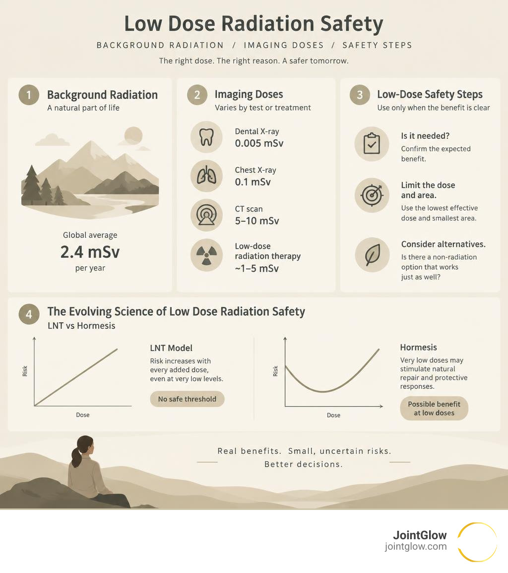
The Evolving Science of Low Dose Radiation Safety: LNT vs Hormesis
For over seven decades, radiation protection standards have been anchored by a single foundational concept: the Linear No-Threshold (LNT) model. This paradigm asserts that biological risk is strictly proportional to dose all the way down to zero. Under LNT, there is no completely “safe” level of ionizing radiation; even a single photon traversing a cell nucleus carries a theoretical risk of causing a malignant transformation.
However, modern radiobiology and clinical research present a much more nuanced picture. A growing body of scientific literature, including comprehensive evaluations such as this scientific review on health impacts of low-dose ionizing radiation, demonstrates that biological organisms possess sophisticated defense mechanisms that respond dynamically to low-level cellular stress (Doss et al., 2014).
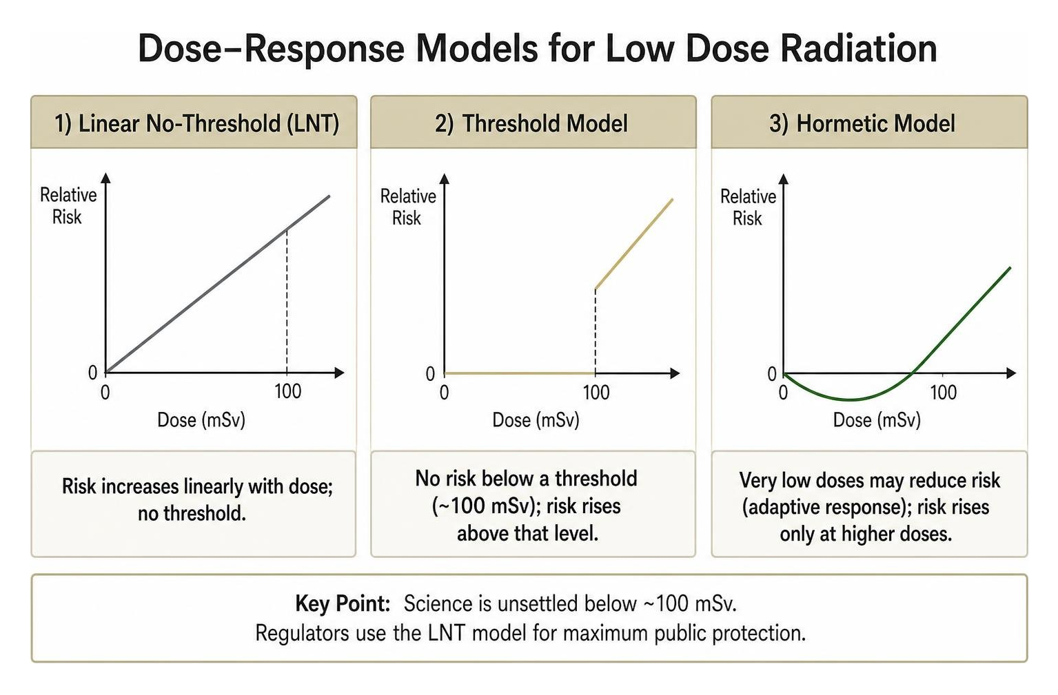
To understand how low dose radiation safety is evaluated today, we must compare the three competing models of radiation dose-response:
| Model | Core Premise | Biological Assumption | Regulatory Status |
|---|---|---|---|
| Linear No-Threshold (LNT) | Risk increases linearly with dose; no exposure level is entirely without risk. | Assumes DNA damage accumulation is linear and repair is imperfect at every scale. | Standard basis for international radiation safety policies (ICRP, NCRP). |
| Threshold Model | Detrimental health effects only occur above a specific absorbed dose (typically ~100 mSv). | Sub-threshold damage is completely repaired by endogenous cellular defense mechanisms. | Supported by numerous clinical and observational cohorts; widely accepted for non-cancer endpoints. |
| Hormesis Model | Ultra-low exposures stimulate protective, anti-inflammatory, and reparative pathways. | Low-level oxidative stress upregulates endogenous antioxidants and systemic immune surveillance. | Demonstrated in experimental biology and specialized therapeutic modalities. |
Historically, LNT was adopted out of regulatory prudence rather than definitive experimental proof at low levels. At very low absorbed doses—defined internationally as 100 milligray (mGy) or less—epidemiological studies face extreme statistical power limitations. When the excess risk is tiny, separating it from the background noise of natural cancer rates, dietary factors, genetics, and lifestyle requires cohorts numbering in the millions. Consequently, while regulators maintain LNT to ensure maximum public precaution, clinical science recognizes that cellular responses to low doses are fundamentally distinct from high-dose tissue destruction.
Biological Mechanisms Informing Low Dose Radiation Safety
At high doses, ionizing radiation overwhelms the cell, causing massive double-strand breaks (DSBs) that lead directly to cell death or severe mutations. At ultra-low doses, however, the biophysical trigger is remarkably precise.
When a low dose of radiation passes through tissue (delivering roughly 1 mGy, which generates DNA DSBs in roughly 3% of exposed cells), the cell activates specialized repair pathways. Scientists monitor this damage using the highly sensitive γ-H2AX foci assay, which detects the phosphorylation of histone variants at the exact site of double-strand breaks. Rather than succumbing to cumulative decay, healthy cells respond by upregulating repair enzymes, stimulating antioxidant production (such as superoxide dismutase and glutathione), and downregulating pro-inflammatory signaling.
This phenomenon encompasses three primary cellular behaviors:
- Adaptive Response: A small initial priming dose “conditions” cells, making them substantially more resistant to subsequent oxidative or radiological stress.
- Bystander Effects: Irradiated cells signal adjacent cells via gap junctions and cytokines, coordinating tissue-level repair and the controlled removal of severely damaged cells through apoptosis.
- Anti-Inflammatory Cascades: Ultra-low doses alter leukocyte adhesion, downregulate pro-inflammatory cytokines like interleukin-1 beta (IL-1β) and tumor necrosis factor-alpha (TNF-α), and shift tissue macrophages from a destructive M1 phenotype toward a reparative, anti-inflammatory M2 phenotype.
Understanding these cellular anti-inflammatory mechanisms allows clinicians to harness ultra-low-dose radiation not as a destructive tool, but as a biological modulator for severe inflammatory conditions.
Evidence Base: Atomic Survivors to Modern Cohorts
Much of what is known about radiation epidemiology originates from the Japanese Life Span Study (LSS), which tracked more than 120,000 survivors of Hiroshima and Nagasaki over multiple decades. The LSS clearly proved the lethality of high-dose radiation: at an acute whole-body dose of 1 Gy, the excess relative risk of solid cancer increases by roughly 50%, while leukemia risk quadruples.
However, the LSS data also show that statistical significance drops off rapidly below 100 mSv. Re-analyses of the data indicate that thresholds for cancer in these cohorts cannot be definitively proven above zero, but neither is an excess risk observable below approximately 60 to 100 mSv. Furthermore, for non-cancer systemic diseases, deterministic injury thresholds appear near 0.9 Sv (900 mSv)—well above any diagnostic or low-dose therapeutic range.
When researchers examine populations living in high natural background radiation areas—such as Ramsar, Iran (where residents receive up to 260 mSv per year) or Kerala, India—they find no statistically significant elevation in cancer mortality or decrease in average lifespan compared to adjacent low-background regions.
Similarly, modern epidemiological restatements, including the comprehensive evidence base restatement on low-level ionizing radiation, note that while pediatric populations show measurable sensitivity to exposures above 5–60 mGy (such as historical multiple-CT cohorts), low-level chronic occupational exposures among nuclear workers often exhibit a “healthy-worker effect” and show no detectable reduction in longevity when modern exposure caps are respected.
ALARA Principles and Practical Radiation Protection
Because science cannot completely rule out stochastic (probabilistic) risks at low levels, the global medical and industrial community operates under the ALARA framework: keeping exposures As Low As Reasonably Achievable, economic and societal factors being taken into account.
ALARA is governed by three classic, universal pillars:
- Time: Minimizing the duration of exposure near any radiation source directly reduces total absorbed dose.
- Distance: Radiation intensity drops off according to the inverse square law. Doubling the distance between an individual and a point source cuts exposure to one-fourth.
- Shielding: Incorporating dense materials—such as lead, bismuth, tungsten, or structural concrete—attenuates ionizing photons before they reach non-target tissues.
Understanding how these pillars function within modern medicine requires precise definitions. In clinical and environmental practice, an absorbed dose under 100 mGy or an effective dose under 100 mSv is categorized as low dose, with dose rates under 0.1 mGy/min classified as low dose-rate. These standard contextual definitions of low-dose radiation ensure that clinical interventions and protective gear are calibrated accurately for safety.

Clinical Guidelines and Occupational Low Dose Radiation Safety
Healthcare workers who perform fluoroscopy-guided interventional procedures—such as interventional cardiologists, radiologists, and orthopedic surgeons—face regular occupational scatter. Protecting these clinicians requires strict procedural workflow management:
- Shielding Configurations: Ceiling-mounted articulated lead-acrylic shields, table-mounted lead skirts, and mobile radiation barrier walls reduce operator exposure by a factor of 20 to 35.
- Beam Geometry: The 90° lateral projection in fluoroscopy generates the highest scatter to the operator (up to ~69 mSv/hour at the tube side), whereas a 45° oblique view yields ~10.9 mSv/hour, and direct anteroposterior (AP) imaging reduces scatter to 4.53–4.98 mSv/hour. Keeping the X-ray tube below the patient and the detector close to the patient dramatically cuts secondary scatter.
- Hardware Adjustments: Using pulsed fluoroscopy (e.g., 7.5 frames per second instead of continuous 30 fps), removing antiscatter grids during non-dense imaging (cutting dose by two- to four-fold), and using tight collimation minimize unnecessary scatter while preserving image clarity.
- Dosimetry Tracking: Real-time electronic dosimeters and monthly film badges worn beneath and outside 0.35–0.5 mm lead-equivalent aprons, thyroid collars, and leaded eyeglasses ensure workers stay well within statutory limits (e.g., 20 mSv/year averaged over 5 years).
Patient Protection Protocols in Modern Medicine
For patients undergoing diagnostic imaging, protection protocols ensure that radiation is delivered with surgical precision:
- Automated Exposure Modulation: Modern CT scanners utilize automated tube-current modulation (adjusting mA in real-time to patient thickness) and advanced iterative reconstruction algorithms to produce diagnostic images at fractions of historical doses.
- Pediatric Image Gently Protocols: Because pediatric tissues divide rapidly and children have longer remaining life spans, pediatric imaging settings strictly scale dose by weight and height, prioritizing non-ionizing alternatives (ultrasound and MRI) whenever clinically indicated.
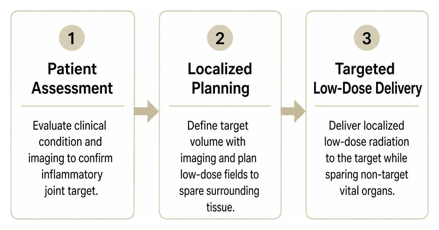
- Targeted Therapeutic Protocols: When low doses are applied therapeutically to treat localized non-malignant conditions, safety relies on modern linear accelerators (LINACs) equipped with multi-leaf collimators. These targeted therapeutic radiation protocols deliver milligray-scale doses exclusively to the inflamed joint space, entirely sparing non-target vital organs.
Therapeutic Low-Dose Radiation: Balancing Risks and Outcomes
While high-dose radiation (typically 40–70 Gy) is utilized in oncology to destroy malignant cells, ultra-low-dose radiotherapy (LDRT) operates in a completely different biological domain. Utilizing localized fractionated doses ranging from 0.5 to 1.0 Gy per session (for a cumulative series well below oncologic thresholds), LDRT targets inflammatory and degenerative musculoskeletal pain.
As documented in historical and contemporary literature, including this scientific re-examination of low-level radiation effects, low-dose treatments do not kill tissue. Instead, they act as a biological switch that disrupts chronic inflammatory cycles:
- Macrophage Reprogramming: Exposure dampens the overproduction of nitric oxide synthase (iNOS) and decreases reactive oxygen species in synovial macrophages.
- Endothelial Modulation: It reduces the adhesion of circulating leukocytes to local vascular endothelium, preventing inflammatory cells from continuously infiltrating the joint.
- Pain Reduction: It halts the local release of pain-mediating neuropeptides, providing durable functional improvement for patients who cannot tolerate or have failed NSAIDs, joint injections, or physical therapy.
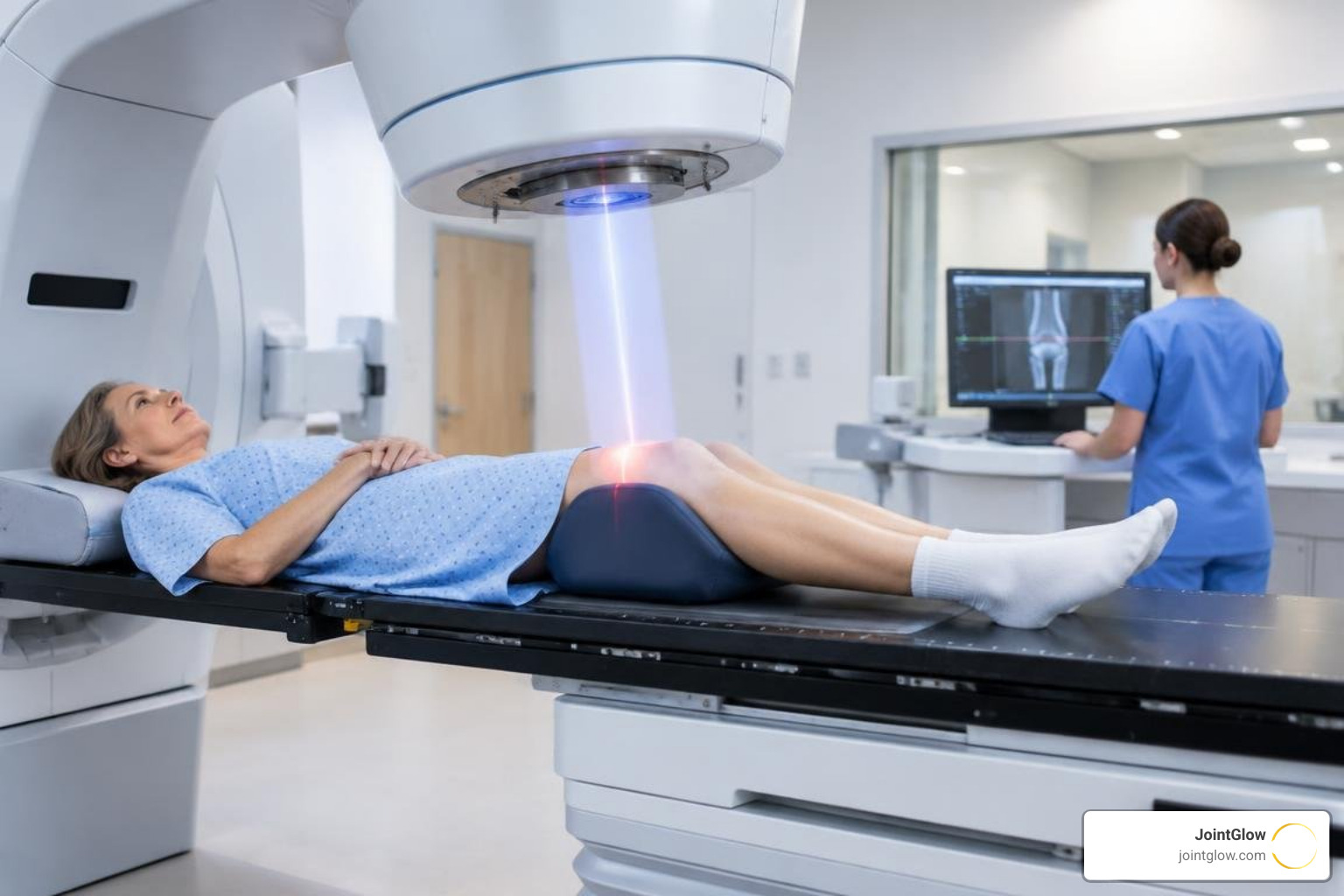
This localized, non-invasive approach is why a growing number of rheumatologists and orthopedic specialists view radiotherapy for chronic osteoarthritis pain as an essential, non-opioid, non-surgical tool for improving joint function and quality of life.
Frequently Asked Questions About Low-Dose Radiation
What is the distinction between stochastic and deterministic effects at low doses?
In radiation biology, health effects are divided into two categories:
- Deterministic Effects (Tissue Reactions): These occur only after reaching a clear physical threshold dose, where a substantial number of cells are damaged or killed. Examples include skin erythema (redness), epilation (hair loss), and radiation-induced cataracts. Below the threshold (typically >500 mGy to several Gy), deterministic injury simply does not occur.
- Stochastic Effects (Probabilistic Risks): These refer to statistical probabilities—primarily cancer induction and hereditary mutations—where the severity of the outcome does not depend on the dose, but the probability of occurrence theoretically increases with dose. Modern low-dose safety guidelines are designed almost entirely to control stochastic probabilities, as diagnostic and low-dose therapies remain orders of magnitude below deterministic injury thresholds.
How does diagnostic imaging exposure compare to natural background radiation?
We live in a naturally radioactive world. Cosmic rays from space, radon gas in the soil, and natural potassium-40 in everyday food (even bananas) contribute to an average worldwide background exposure of ~2.4 mSv per year (roughly 0.01 mSv/day). In some regions, background levels naturally reach 10 to 50 mSv annually without adverse population health effects.
Putting common medical procedures into context:
- Chest X-ray: ~0.02 to 0.1 mSv (equivalent to a few days of natural background radiation).
- Mammogram: ~0.4 mSv (equivalent to ~7 weeks of background exposure).
- Head CT: ~1.5 to 2.0 mSv (equivalent to ~8 to 10 months of background exposure).
- Abdominal/Pelvic CT: ~7.0 to 10.0 mSv (equivalent to ~3 to 4 years of natural background radiation).
When diagnostic imaging is medically indicated, the immediate diagnostic benefit dramatically outweighs the infinitesimal statistical risk associated with a few months or years of background radiation equivalent.
Does current scientific evidence support a clear safety threshold below 100 mSv?
Direct epidemiological data have consistently failed to demonstrate statistically significant increases in cancer incidence or mortality at acute whole-body exposures below 100 mSv (and chronic cumulative exposures far higher). While laboratory models confirm that single-track radiation causes DNA double-strand breaks at very low doses (1 mGy), human tissues possess highly efficient enzymatic DNA repair mechanisms that correct these baseline breaks in healthy cells.
International bodies such as the ICRP and UNSCEAR maintain conservative linear risk estimates for public protection policies, but consensus panels widely acknowledge that below 100 mSv, potential health risks are so small that they are effectively indistinguishable from zero against standard baseline cancer rates.
Conclusion
The debate surrounding low-dose ionizing radiation has evolved from reflexive radiophobia toward an evidence-based understanding of biological response. While the ALARA principle remains an indispensable safeguard against careless or unnecessary exposure, clinical data confirm that low-dose radiation behaves uniquely at the cellular level—triggering DNA repair, mitigating oxidative stress, and dampening chronic inflammatory responses.
At JointGlow, we leverage this advanced radiobiological science to deliver precise, localized, Medicare-covered Low-Dose Radiotherapy (LDRT) for individuals suffering from chronic, debilitating joint pain. By delivering non-invasive, targeted low doses directly to inflamed joints, our therapy resets dysfunctional cellular inflammation without surgery, anesthesia, systemic medications, or downtime.
If you are exploring non-surgical solutions for severe osteoarthritis or chronic joint inflammation, consult our medical team and review the expected timeline for therapeutic relief to see how precision low-dose therapy can help restore your mobility.
Scientific References
Doss M, Little MP, Orton CG. “Point/Counterpoint: low-dose radiation is beneficial, not harmful..” Medical physics, 2014. PMCID PMC4109571.
