First-Line Medial Epicondylitis Treatment and At-Home Management
When treating golfer’s elbow, our primary goal is to offload the common flexor tendon origin so normal tissue remodeling can occur. Because up to 90% of cases stem from occupational tasks rather than sports, first-line management centers on practical load management, temporary splinting, and structured movement. For a thorough overview of these non-operative principles, see this evidence-based overview of medial epicondylitis.
Initial Rest, Cryotherapy, and Counterforce Bracing
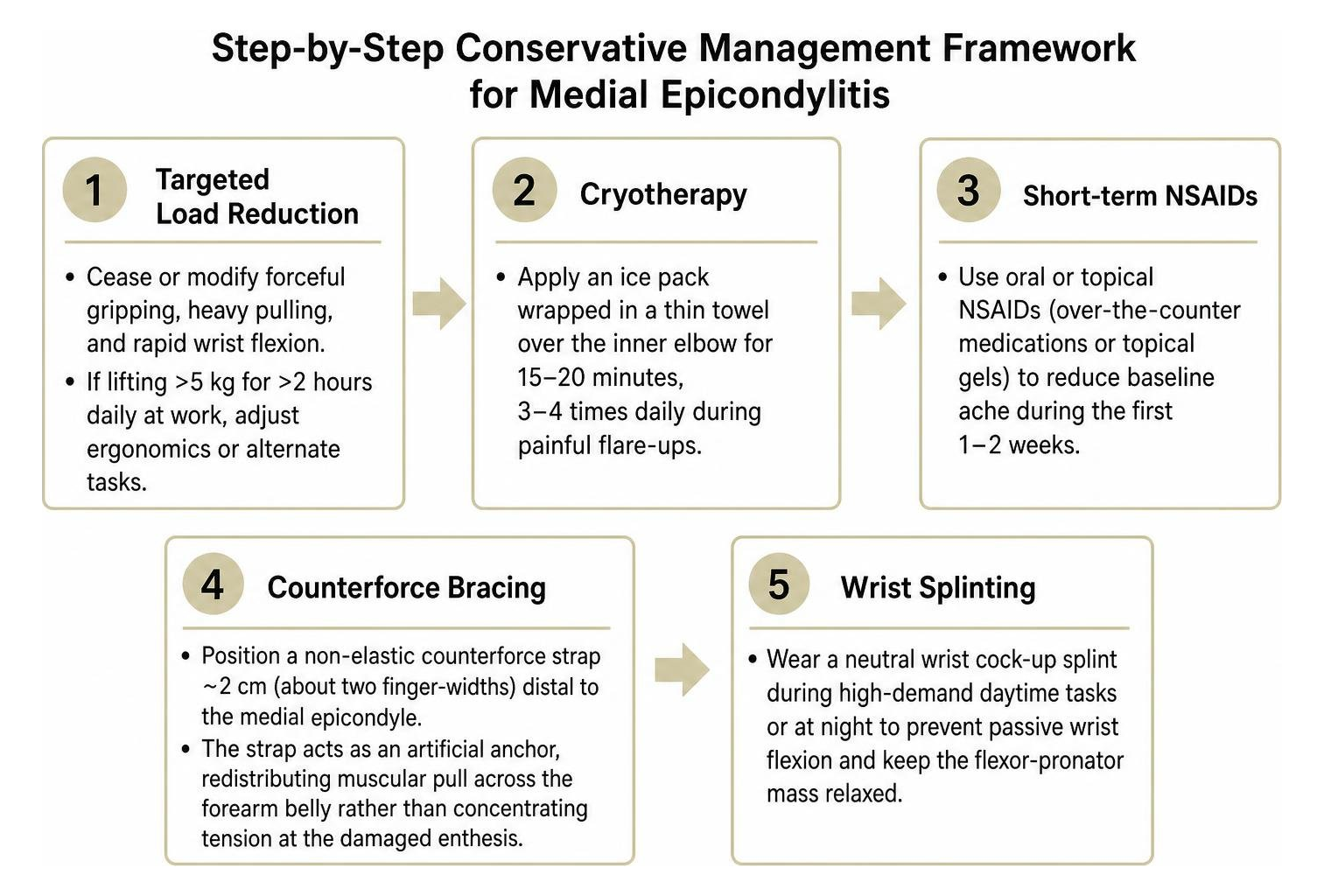
Early conservative management focuses on dampening acute symptoms without completely immobilizing the arm:
- Targeted load reduction: Cease or modify forceful gripping, heavy pulling, and rapid wrist flexion. If your job involves lifting items heavier than 5 kg for more than 2 hours per day, adjust your ergonomics or alternate tasks to prevent microtrauma accumulation.
- Cryotherapy: Apply an ice pack wrapped in a thin towel directly over the inner elbow for 15 to 20 minutes, 3 to 4 times daily during painful flare-ups.
- Short-term oral or topical NSAIDs: Over-the-counter anti-inflammatory medications or topical gels can alleviate baseline ache during the first 1 to 2 weeks.
- Counterforce bracing: Position a non-elastic counterforce strap approximately 2 cm (about two finger-widths) distal to the medial epicondyle. This strap acts as an artificial anchor, redistributing muscular pull across the forearm belly rather than concentrating mechanical tension at the damaged enthesis.
- Wrist splinting: A neutral wrist cock-up splint worn during high-demand daytime tasks or at night prevents passive wrist flexion, keeping the flexor-pronator mass relaxed.
Progressive Physical Therapy and Eccentric Strengthening
Once the acute resting ache subsides, true tendon recovery requires mechanical loading. Tendons require controlled tensile stimulus to align new collagen fibers. Clinicians recommend following a structured progression as outlined in this clinical guide to epicondylitis management:
- Passive Wrist Flexor Stretching: Extend the elbow fully with the palm facing up. Use your opposite hand to gently pull the wrist and fingers backward into extension until a gentle stretch is felt along the inner forearm. Hold for 20 to 30 seconds, repeating 3 to 4 times daily.
- Isometric Loading: With the forearm supported on a flat table, hold the wrist in a neutral posture against static manual resistance without letting the joint move. Hold for 10 to 15 seconds, resting between repetitions.
- Eccentric Flexor-Pronator Strengthening: Hold a light weight (such as a 1 to 2 lb dumbbell or a soup can). Use the unaffected hand to lift the wrist into flexion (the concentric phase), then slowly lower the weight downward into extension over a strict 4-second count (the eccentric phase). Perform 3 sets of 10 to 15 repetitions daily.
- Therapy Putty and Grip Work: Squeeze low-resistance therapy putty or a rolled towel, focusing on controlled finger flexion and abduction.
Non-Invasive Treatments for Chronic Medial Elbow Pain
For the 10% to 15% of individuals whose symptoms do not resolve after 6 to 12 weeks of standard physical therapy and load modification, progressing to advanced non-surgical modalities is the next logical step. Chronic cases shift from a simple strain into a persistent degenerative cycle marked by hypervascularity, localized tissue thickening, and neurogenic sensitization. Exploring advanced non-surgical joint treatments helps address this chronic tissue change without surgical risks.
Low-Dose Radiotherapy for Persistent Medial Epicondylitis
When chronic inflammation and hypervascular remodeling become fixed, standard modalities may reach a plateau. Low-dose radiotherapy (LDRT) is an established, non-invasive therapeutic modality utilized widely for benign hyperproliferative and chronic inflammatory musculoskeletal disorders.
LDRT delivers ultra-low-dose radiation fractions (substantially lower than oncologic doses) to the medial epicondyle. At these calibrated fractions, the treatment works at the cellular level by:
- Decreasing inflammatory macrophage adhesion and nitric oxide production.
- Disrupting pathologic neovascular networks that carry nociceptive (pain-sensing) nerve fibers.
- Restoring local microenvironmental balance to promote durable tissue calm.
Because LDRT involves no needles, incisions, or rehabilitation downtime, it offers a compelling option for eligible patients—and is covered by Medicare Part B (Excludes Medicare Advantage) for qualifying chronic conditions.
Advanced Non-Invasive Care for Chronic Tendon Pain

For individuals experiencing long-standing tendon pain, non-invasive clinical protocols focus on resetting persistent inflammatory cycles and encouraging functional recovery:
- Targeted Anti-Inflammatory Modulation: Cellular-level therapies help calm localized cytokine cascades, creating an environment favorable for tissue stabilization.
- Neuromodulation of Sensitized Pathways: By addressing neurogenic sensitization, non-invasive modalities dampen overactive pain signaling around the medial epicondyle.
- Preservation of Structural Integrity: Non-invasive care avoids tissue trauma, injections, or prolonged immobilization, facilitating an active return to daily activities and rehabilitation.
When to Seek Specialist Evaluation for Persistent Symptoms
While over 85% to 90% of patients successfully recover with non-operative care within several months, persistent symptoms warrant specialist assessment to prevent long-term joint dysfunction (NBK557869). The StatPearls review of medial epicondylitis emphasizes that early diagnostic clarification ensures rare complications are addressed before permanent structural changes occur (NBK557869).
Evaluation Criteria for Refractory Medial Epicondylitis
Consult an orthopedic or sports medicine specialist if you observe any of the following benchmarks:
- Symptoms persist beyond 3 to 6 months despite consistent, structured physical therapy, activity adjustment, and bracing (Konarski et al., 2023).
- Progressive loss of forearm grip strength interfering with basic activities of daily living (e.g., turning doorknobs, opening jars).
- Night pain or resting ache that disrupts sleep.
- Inability to perform occupational duties without sharp, debilitating medial elbow discomfort.
Addressing Concomitant Ulnar Nerve Neuropathy
An essential reason to seek specialist care is the anatomical proximity of the ulnar nerve, which passes through the cubital tunnel directly posterior to the medial epicondyle (NBK557869).
Up to 20% to 60% of individuals with chronic medial epicondylitis demonstrate concomitant ulnar neuritis or cubital tunnel syndrome (NBK557869).
When ulnar neuritis is present, injections near the epicondyle must be performed with extreme care under direct ultrasound visualization to avoid direct intraneural injection (NBK551506).
Accurately Diagnosing Medial Elbow Pain and Differential Conditions
A thorough diagnostic workup begins with a physical examination to isolate the exact source of pain, as outlined in the Merck Manual clinical evaluation of medial epicondylitis.
Physical Provocation Tests and Diagnostic Imaging
Clinical diagnosis relies on reproducible provocative testing:
- Point Tenderness: Maximal focal tenderness is typically isolated approximately 5 to 10 mm distal and anterior to the medial epicondyle, directly over the pronator teres and flexor carpi radialis origins (NBK557869).
- Resisted Wrist Flexion Test: With the patient’s elbow extended and forearm supinated, the examiner resists active wrist flexion. A sharp reproduction of pain at the medial epicondyle is a positive indicator (NBK557869).
- Resisted Forearm Pronation: Resisting active pronation with the elbow positioned at 90 degrees stresses the pronator teres origin, eliciting medial elbow discomfort (NBK557869).
When symptoms are atypical or persistent, imaging clarifies the diagnosis:
- Musculoskeletal Ultrasound: Highly accessible with diagnostic sensitivity exceeding 95% and specificity around 92% (NBK557869). It demonstrates common flexor tendon thickening, hypoechoic degenerative zones, intrasubstance tears, and calcifications (Konarski et al., 2023).
- Magnetic Resonance Imaging (MRI): The gold standard for evaluating tendon architecture, intra-articular pathology, and soft-tissue changes (NBK557869).
- Radiographs (X-rays): Primarily used to exclude loose bodies, traction spurs, or medial compartment osteoarthritis (NBK557869).
Differentiating Tendinopathy from Ligament and Joint Pathologies
Inner elbow pain is not always golfer’s elbow. To ensure successful recovery, clinicians distinguish tendinopathy from other medial joint issues:
| Differential Diagnosis | Key Distinguishing Features | Diagnostic Maneuver / Test |
|---|---|---|
| Medial Epicondylitis | Pain localized 5–10 mm distal to epicondyle; intact joint stability (NBK557869) | Pain on resisted wrist flexion & pronation (NBK557869) |
| UCL Insufficiency | Common in throwing athletes; medial joint instability | Positive Moving Valgus Stress Test; Milking Maneuver |
| Cubital Tunnel Syndrome | Paresthesias in 4th/5th digits; grip weakness | Positive Tinel test at cubital tunnel; EMG/NCS |
| Medial Joint Osteoarthritis | Joint line stiffness, crepitus, loss of terminal extension | Radiographs showing joint space narrowing and osteophytes |
| Cervical Radiculopathy (C8-T1) | Neck pain radiating down medial forearm; dermatomal deficit | Spurling test; cervical spine MRI |
For patients navigating complex or long-standing joint discomfort, managing chronic elbow conditions with comprehensive diagnostic evaluation ensures that secondary issues like joint wear or nerve irritation are addressed.
Frequently Asked Questions About Golfer’s Elbow
For more practical guidance on recovery milestones, visit our collection of frequently asked recovery questions.
How long does it take for golfer’s elbow to fully heal?
The natural recovery timeline varies according to chronicity:
- Acute Cases (symptoms < 6 weeks): Typically improve within 6 to 12 weeks of consistent activity modification, bracing, and progressive home exercises.
- Chronic Degenerative Cases (symptoms > 3 months): Often require 6 to 12 months of structured physical therapy, eccentric loading, or advanced non-invasive therapies to remodel degenerative collagen tissue (NBK557869, Konarski et al., 2023).
- Surgical Recovery: If open or arthroscopic debridement is performed, full return to high-demand athletic activities or heavy manual labor takes approximately 4 to 6 months post-operatively.
Why can persistent medial epicondylitis require a different treatment approach?
Persistent medial epicondylitis is pathologically characterized by angiofibroblastic tendinosis—a state of disorganized collagen repair, microtears, and abnormal ingrowth of sensory nerves and small vessels—rather than active inflammatory cellular infiltration (NBK557869, Konarski et al., 2023). Consequently, treatments aimed solely at masking acute inflammation (such as repeated cortisone injections) fail to stimulate long-term structural repair and can weaken collagen integrity. Chronic cases benefit most from targeted biological reset strategies, including structured eccentric loading programs and LDRT (NBK551506, Konarski et al., 2023).
What daily habits or occupational factors worsen medial elbow pain?
The primary contributors to recurrent tendon strain include:
- Repetitive Wrist Flexion Under Load: Heavy manual tasks such as bricklaying, plumbing, carpentry, and continuous hammering.
- Improper Sports Mechanics: Leading with an open elbow during the golf downswing (“hitting from the top”), throwing sidearm with excessive valgus stress, or using a tennis racket with an undersized grip.
- Poor Ergonomics: Prolonged typing on non-ergonomic keyboards with wrists resting in extreme flexion or pronation without forearm support.
- Systemic Risk Factors: Elevated body mass index (BMI), diabetes mellitus, and smoking, which impair localized microvascular blood supply and delay soft-tissue healing.
Conclusion
Overcoming medial epicondylitis requires a stepwise, evidence-based approach. By recognizing that golfer’s elbow is a degenerative tendinopathy rather than an acute inflammatory episode, you can focus on therapies that restore tissue health: load management, counterforce bracing, and progressive eccentric strengthening (NBK557869, Konarski et al., 2023).
When standard home routines stall, non-invasive therapeutic options such as low-dose radiotherapy (LDRT) offer a drug-free, non-surgical path to reset persistent discomfort. If you are struggling with persistent inner elbow pain that interferes with your daily activities or work, discover our approach to specialized care for chronic elbow pain and begin your journey toward lasting relief today.
References
NBK557869. NBK551506. Konarski W, Poboży T, Poboży K, Domańska J, Konarska K. “Current concepts of natural course and in management of medial epicondylitis: a clinical overview..” Orthopedic reviews, 2023. PMCID PMC10495044.
