Joint Inflammation: What It Means for Your Pain
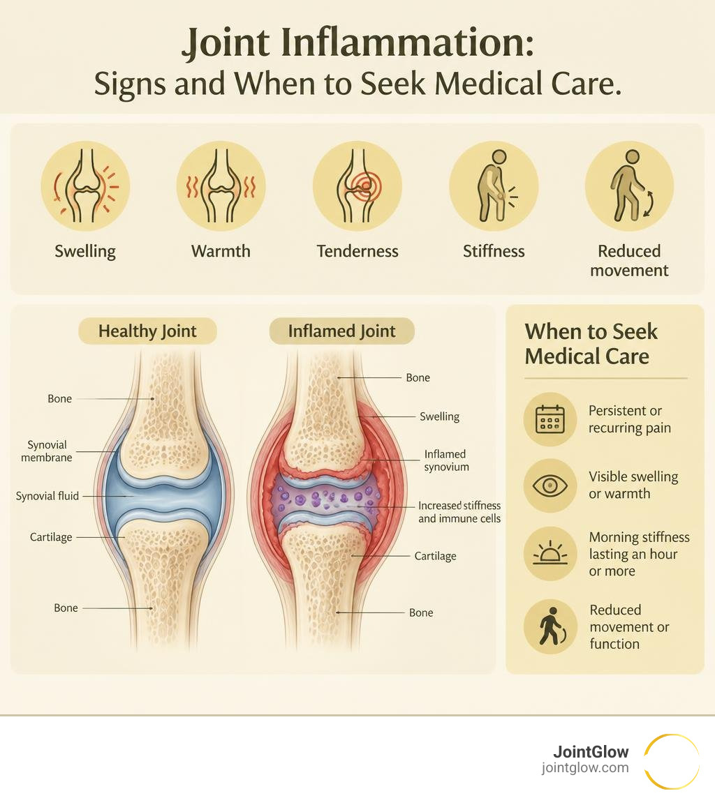
Joint inflammation happens when the lining of a joint or nearby tissues react to injury, crystal buildup, infection, or immune-system activity. It can cause swelling, warmth, tenderness, stiffness, and reduced movement.
A brief flare after a strain may improve with rest and gentle care. But pain that keeps coming back – especially with visible swelling, warmth, or morning stiffness lasting an hour or more – can point to inflammatory arthritis and deserves medical evaluation. Early treatment matters because ongoing inflammation can damage cartilage, bone, ligaments, and the joint itself.
Joint pain is not always inflammation. Wear-and-tear osteoarthritis often gets worse with use and may cause grinding or creaking. Inflammatory pain is more likely to hurt at rest, affect several joints, and ease somewhat as you start moving.
In this guide, I will help you understand the possible causes, warning signs, testing, and treatment options so you can have a more informed conversation with your care team. I am Dr. Koneru, and we will start with what is happening inside an inflamed joint.
What Is Joint Inflammation and How Does It Develop?
To understand how joint inflammation develops, we have to look closely at synovial joints—the flexible hinges and pivots like your knees, hips, hands, and shoulders. Under normal circumstances, a smooth layer of cartilage cushions the ends of your bones, and a thin, delicate lining called the synovial membrane (or synovium) encapsulates the space. This membrane produces a tiny amount of synovial fluid that lubricates movement, much like motor oil in an engine.
When an irritant enters the picture—whether a sports injury, sharp microscopic crystals, or misdirected immune cells—the body initiates a rapid defense sequence. Blood vessels in the joint capsule dilate, causing increased vascular permeability. White blood cells, plasma fluids, and inflammatory mediators flood into the enclosed synovial cavity.
This leads directly to synovitis (inflammation of the synovial lining) and joint effusion, an abnormal buildup of fluid that creates internal pressure, visible swelling, and that familiar tight, throbbing ache. Over time, persistent immune activation changes the synovial environment from protective to destructive. As detailed in research on chronic inflammation and joint tissue degradation, chronic inflammatory signaling causes the synovium to thicken and release enzymes that progressively break down underlying cartilage (NBK507704).
Acute vs Chronic Joint Inflammation
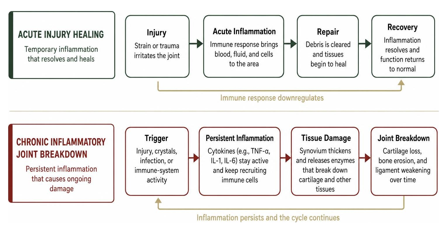
Not all inflammation is bad. In fact, acute inflammation is a vital, healthy biological response. If you twist your ankle stepping off a curb, acute inflammation immediately rushes blood, nutrients, and immune cells to the scene to stabilize tissues, clear cellular debris, and begin tissue repair. Once trauma recovery is underway, the immune system downregulates itself, the swelling subsides, and joint function returns to normal.
Chronic joint inflammation, however, is a destructive biological loop. Instead of shutting down after tissue repair is complete, cytokine cascades (involving inflammatory proteins like TNF-alpha, IL-1, and IL-6) remain constantly active. This persistent state fails to heal the joint; instead, it recruits more immune cells, creating an ongoing attack that damages cartilage, weakens stabilizing ligaments, and erodes bone surfaces over months and years.
Cellular Pathways and the Autoimmune Attack
In autoimmune forms of arthritis, the immune system mistakes healthy joint structures for foreign invaders. Circulating leukocytes (white blood cells, including T cells and B cells) cross through permeable blood vessel walls and infiltrate the synovium.
As these immune cells accumulate, they produce localized antibodies, recruit destructive macrophages, and promote neovascularization (the growth of abnormal, fragile new blood vessels). This hyperactive, thickened synovial tissue transforms into an invasive mass called a pannus. The pannus creeps over articular cartilage, releasing matrix metalloproteinases and other aggressive enzymes that eat away at both cartilage and subchondral bone.
Understanding these cellular mechanisms of inflammatory signaling is what allows modern medicine to design targeted therapies that halt tissue degradation at the microscopic level.
Primary Causes of Inflammatory Arthritis and Swelling
Inflammatory arthritis accounts for an estimated annual incidence of 115 to 271 cases per 100,000 adults. In the United States, an estimated 22.7% of all adults live with some diagnosed form of arthritis. Identifying what drives inflammatory swelling is crucial, as causes range from systemic autoimmunity to localized metabolic imbalances.
A comprehensive clinical evaluation of polyarticular joint pain typically categorizes systemic inflammatory arthritis into several distinct conditions:
- Rheumatoid Arthritis (RA): The most common autoimmune inflammatory arthritis worldwide. RA classically causes symmetrical joint inflammation, targeting the small joints of the hands, wrists, and feet, accompanied by severe morning stiffness and fatigue.
- Psoriatic Arthritis (PsA): About 30% of people with the skin condition psoriasis develop psoriatic arthritis. PsA can cause asymmetric joint inflammation, dactylitis (“sausage digits”), spine stiffness, and nail changes.
- Ankylosing Spondylitis and Axial Spondyloarthritis: Inflammatory conditions strongly associated with the genetic marker HLA-B27, primarily targeting the sacroiliac joints and spine, leading to chronic back stiffness and potential spinal fusion if untreated.
- Systemic Lupus Erythematosus (SLE): A multi-system autoimmune disease that frequently triggers migratory, non-erosive polyarthritis alongside skin rashes and kidney or heart inflammation.
- Infectious (Septic) Arthritis: A medical emergency where bacteria (such as Staphylococcus aureus) or viruses directly enter the joint space, triggering massive, rapid-onset joint effusion and severe pain.
Crystal-Induced Arthropathies and Gout
Not all inflammatory joint disease stems from autoimmunity. Crystal arthropathies occur when mineral deposits accumulate directly inside the joint space, triggering sudden, agonizing inflammatory flares.
Gout is the single most common form of inflammatory arthritis overall. It occurs when high levels of serum uric acid precipitate out of the bloodstream and form needle-sharp monosodium urate crystals within the joint fluid. White blood cells ingest these crystals, triggering an explosive inflammatory surge. Roughly 50% of gout patients present with podagra—a classic, excruciating attack localized to the first metatarsophalangeal (MTP) joint of the big toe. Over time, untreated hyperuricemia leads to firm, chalky nodular deposits known as tophi, which can erode nearby bone and permanently deform the joint.
A related condition, Calcium Pyrophosphate Deposition (CPPD) disease—sometimes referred to as pseudogout—occurs when calcium pyrophosphate dihydrate crystals settle into cartilage, frequently triggering sudden, hot, swollen attacks in the knees or wrists (NBK507704). A detailed overview of crystalline and inflammatory arthropathies highlights how crystal identification under polarized light microscopy provides a definitive diagnostic answer.
Periarticular Irritation and Soft Tissue Conditions
Sometimes, severe pain that feels like deep joint inflammation actually originates in the soft tissues surrounding the joint capsule. Periarticular structures include tendons (which connect muscle to bone) and bursae (small, fluid-filled sacs that reduce friction between bones and soft tissue).
Repetitive strain, sudden impact, or biomechanical imbalances can cause acute tendinitis or severe periarticular swelling from bursitis. When these tissues inflame, movement becomes painful, often mimicking true intra-articular arthritis. Distinguishing between deeper joint capsule swelling and superficial soft tissue irritation is an essential part of diagnosing complex inflammatory joint conditions.
Inflammatory Arthritis vs. Osteoarthritis: Key Diagnostic Differences
One of the most frequent questions we encounter in clinical practice is how to tell whether joint discomfort is due to mechanical “wear-and-tear” or underlying systemic inflammation.
While modern research shows that low-grade inflammation can accompany mechanical osteoarthritis, the core drivers, clinical presentations, and treatment pathways are fundamentally different. Understanding how to distinguish mechanical joint pain from systemic inflammation is critical for preventing irreversible joint damage:
| Clinical Feature | Osteoarthritis (Mechanical) | Inflammatory Arthritis (Systemic / Autoimmune) |
|---|---|---|
| Primary Cause | Cartilage wear, aging, mechanical overload | Immune system dysfunction, crystal deposition, infection |
| Morning Stiffness | Brief; typically lasts under 30 minutes | Prolonged; commonly lasts over 60 minutes |
| Pain Pattern | Worsens with physical activity; eases with rest | Constant or worse at rest; improves with gentle movement |
| Joint Appearance | Bony enlargement (Heberden’s nodes), cool to touch | Visible swelling, warmth, redness (erythema), soft effusion |
| Symmetry | Often asymmetrical (e.g., one knee, one hip) | Frequently symmetrical (e.g., matching knuckles on both hands) |
| Audible Cues | Common crepitus (grating, creaking sounds) | Less common; primary feature is fluid tension and tightness |
| Systemic Signs | None (localized to joint) | Frequent fatigue, fever, skin rashes, eye inflammation |
Clinical Evaluation and Treatment Pathways
Getting an accurate diagnosis requires a structured, multi-step clinical evaluation. Because undifferentiated inflammatory arthritis progresses to rheumatoid arthritis in roughly 13% to 54% of cases, catching the disease early gives patients the best chance of preserving normal joint anatomy.
A comprehensive diagnostic workup includes:
- Detailed Clinical History & Physical Exam: Evaluating the number of involved joints, checking for warmth and soft tissue effusion, and testing both active and passive range of motion.
- Inflammatory Blood Markers: Testing for elevated Erythrocyte Sedimentation Rate (ESR) and C-Reactive Protein (CRP), which indicate systemic immune activity.
- Autoantibody Screening: Checking for Rheumatoid Factor (RF), anti-cyclic citrullinated peptide (anti-CCP), and Antinuclear Antibodies (ANA) to detect autoimmune patterns.
- Synovial Fluid Analysis (Arthrocentesis): Aspiration of joint fluid to assess white blood cell counts (counts above 5,000 cells/mm³ suggest inflammatory arthritis, while counts above 50,000 cells/mm³ strongly indicate septic arthritis) and analyze crystal morphology under microscopy.
- Diagnostic Imaging: Plain radiographs detect late-stage joint space narrowing and bone erosion, while musculoskeletal ultrasound and MRI visualize early synovitis, joint effusion, and soft tissue changes before permanent damage occurs.

Once diagnosed, managing inflammatory pain involves combining evidence-based medications, physical therapy, and innovative therapeutic interventions. Our full spectrum of evidence-based joint treatments includes conventional disease-modifying antirheumatic drugs (DMARDs), targeted biologic therapies, Janus kinase (JAK) inhibitors, and non-pharmacologic modalities.
Complications of Untreated Joint Inflammation
Ignoring chronic joint swelling is risky. When joint tissues remain chronically inflamed, enzymes and cellular pressure steadily dismantle structural anatomy.
Long-term, untreated inflammatory arthritis can lead to severe joint subluxation (partial dislocation), profound cartilage loss, and irreversible ankylosis (bony or fibrous joint fusion). Beyond local joint deformity and permanent functional disability, uncontrolled systemic inflammation increases cardiovascular risks, contributes to osteoporosis, and causes persistent centralized pain sensitivity. Accessing thorough patient educational resources early helps individuals take control of their joint health before structural changes become permanent.
Low-Dose Radiotherapy for Joint Inflammation
For patients seeking non-invasive, drug-free relief from persistent, painful joint inflammation, Low-Dose Radiotherapy (LDRT) represents a clinically proven treatment pathway. Widely utilized in European clinics for decades and fully covered by Medicare Part B (Excludes Medicare Advantage) in the United States, LDRT uses an ultra-low, precisely targeted dose of ionizing radiation—only a tiny fraction of the doses used in oncology—to calm inflamed tissues.
At the cellular level, LDRT alters the phenotype of hyperactive immune cells, downregulates pro-inflammatory cytokines, diminishes vascular permeability, and encourages macrophage clearance. This biologically resets joint inflammation, breaking the chronic cycle of pain and swelling without surgery, injections, systemic drug toxicity, or downtime. Discover the full details on how low-dose radiotherapy works to provide durable, restorative pain relief.
Frequently Asked Questions About Inflamed Joints
Can diet and exercise reduce swelling in affected joints?
Yes, lifestyle choices play a significant supportive role in controlling systemic inflammation. Following an anti-inflammatory nutritional pattern—such as the Mediterranean diet, rich in extra virgin olive oil, leafy greens, berries, nuts, and omega-3 fatty acids from fatty fish—helps reduce circulating inflammatory markers.
Pairing nutrition with low-impact aerobic exercise (such as swimming, cycling, or walking) lubricates joints, strengthens supporting musculature, and prevents stiffness. Weight loss also substantially decreases mechanical load on weight-bearing joints while reducing pro-inflammatory adipokines produced by visceral fat tissue.
When should you see a doctor for swollen, painful joints?
You should seek prompt medical evaluation if you experience any of the following “red flag” symptoms:
- A joint that is suddenly hot to the touch, visibly bright red, and severely swollen.
- Joint pain accompanied by a fever, chills, or unexplained weight loss.
- Sudden inability to bear weight or move the joint.
- Severe morning stiffness that lasts longer than an hour.
- Joint swelling accompanied by new rashes, eye pain, or mouth ulcers.
These symptoms warrant immediate assessment to rule out septic arthritis, acute crystalline gout flares, or aggressive new-onset autoimmune disease.
Can joint inflammation resolve entirely on its own?
Mild, acute inflammation following a minor strain or overexertion can resolve on its own with temporary rest, ice, elevation, and gentle activity modification.
However, chronic or recurrent inflammation caused by autoimmune conditions, persistent crystal buildup, or structural joint breakdown will not disappear spontaneously. Leaving these conditions unaddressed allows inflammatory enzymes to steadily erode cartilage and bone beneath the surface. Early professional intervention is essential to protect joint longevity.
Conclusion
Joint inflammation is your body’s distress signal. Whether triggered by an acute mechanical injury, crystalline deposits, or a complex autoimmune response, persistent joint swelling and prolonged morning stiffness should never be ignored. Early diagnosis is the cornerstone of joint preservation, preventing irreversible bone erosions and safeguarding your long-term mobility.
At JointGlow, we are dedicated to helping you overcome debilitating joint pain and regain an active, unrestricted life through advanced, non-invasive therapies. If you are exploring non-surgical options to calm chronic joint inflammation, review our comprehensive timeline for non-invasive joint pain relief to see when you can expect meaningful, long-lasting comfort.
