Why Ankylosing Spondylitis Inflammation Can Cause Lasting Spinal Pain
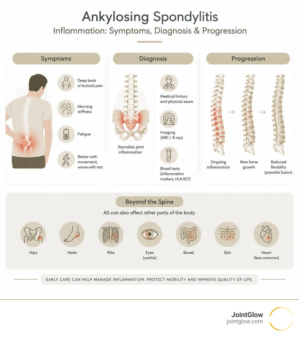
Ankylosing spondylitis inflammation is an overactive immune response that mainly attacks the sacroiliac joints and spine. It can cause deep low-back or buttock pain, morning stiffness, fatigue, and pain that often improves after moving but worsens with rest. Over time, repeated inflammation may trigger abnormal new bone growth, reducing flexibility and, in some people, fusing parts of the spine.
This is more than ordinary wear-and-tear back pain. Ankylosing spondylitis is part of axial spondyloarthritis, a chronic inflammatory disease that can also affect the hips, heels, ribs, eyes, bowel, skin, and, less often, the heart.
Early care matters. A rheumatologist can assess persistent back pain that began gradually, wakes you at night, or comes with prolonged morning stiffness. Sudden red, painful eyes or vision changes need urgent medical attention because they may signal uveitis.
In the sections ahead, I am Dr. Koneru, and I will explain what drives this inflammation, how it leads to spinal damage, and the options used to protect mobility and manage pain.
What Causes Ankylosing Spondylitis Inflammation: Genetics, Microbiome, and Cellular Triggers
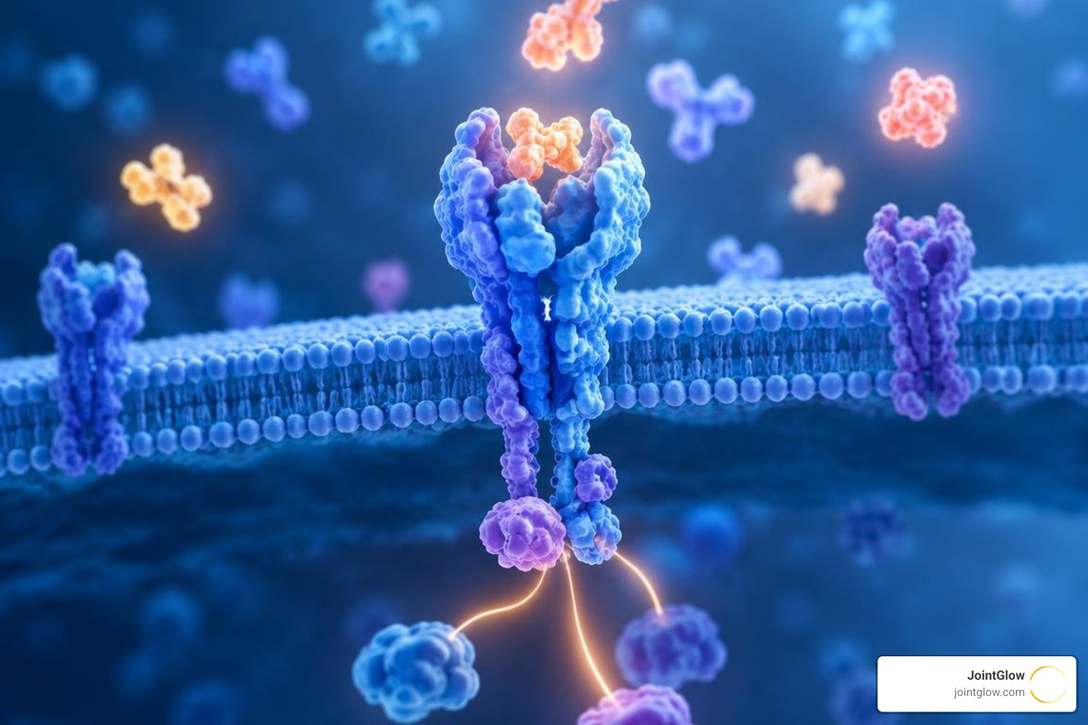
Ankylosing spondylitis (AS) does not happen in a vacuum. It sits at the intersection of our genetics, our environment, and deep cellular immune signaling. While the condition was historically categorized as a simple autoimmune disease, modern clinical research reveals that it functions as a complex hybrid of autoinflammatory and autoimmune mechanisms.
To understand why inflammation takes hold, researchers have turned their attention toward genetic variations, intestinal barrier disruptions, and the inner mechanics of our cells. Breakthrough pathogenesis research in axial spondyloarthritis has uncovered that the disease originates from multiple biological checkpoints failing simultaneously. When these barriers break down, the body unleashes persistent, localized attacks on joint tissues.
The Role of HLA-B27 and Genetic Predisposition
The most prominent genetic risk factor linked to axial spondyloarthritis is Human Leukocyte Antigen B27 (HLA-B27). This major histocompatibility complex (MHC) class I molecule helps our immune systems tell the difference between healthy self-proteins and foreign invaders like bacteria or viruses.
However, HLA-B27 has a structural quirk: its heavy chains are unusually prone to misfolding inside the cell’s protein factory, known as the endoplasmic reticulum (ER). When misfolded proteins accumulate, they trigger intense ER stress and activate the unfolded protein response (UPR). This internal cellular distress signal prompts immune cells to release high volumes of pro-inflammatory cytokines, especially interleukin-23 (IL-23).
Along with HLA-B27, genetic variants in the endoplasmic reticulum aminopeptidase (ERAP1 and ERAP2) genes alter how peptides are trimmed and presented to immune cells. Furthermore, research highlights a vital gut-joint axis. Many people with AS carry an altered microbiome—a state known as gut dysbiosis—often marked by an abundance of specific microbes such as Klebsiella pneumoniae. When the gut lining becomes permeable, microbial fragments cross into circulation, prompting immune cells to travel directly from the digestive tract to spinal attachment sites.
Cellular Mechanisms Fueling Ankylosing Spondylitis Inflammation
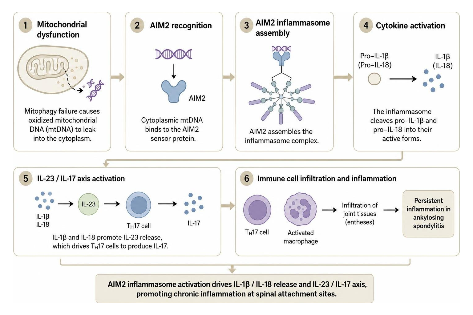
Beyond genetic susceptibility, inflammation is sustained by cellular stress pathways. An important modern discovery is the link between damaged cell powerhouses (mitochondria) and persistent immune activation. A comprehensive mitochondrial dysfunction and AIM2 inflammasome review explains how oxidized mitochondrial DNA (mtDNA) leaks into the cytoplasm of immune cells when cellular recycling (mitophagy) fails.
Once in the cytoplasm, this free-floating mtDNA acts as a danger signal, binding directly to the AIM2 (Absent in Melanoma 2) sensor protein. This binding triggers the assembly of the AIM2 inflammasome, which converts inactive pro-interleukins into mature, destructive IL-1β and IL-18.
At the same time, this activation feeds the powerful IL-23/IL-17 cytokine axis, driving T helper 17 (TH17) cells and specialized macrophages to infiltrate joint capsules. Recent NK-cell antigen presentation analysis demonstrates that natural killer cells can also switch roles to present antigens directly via HLA-DPB1/DPA1 pathways, further escalating T-cell activity. At JointGlow, exploring the science of inflammation at the cellular level is foundational to how we understand joint degeneration and develop targeted interventions.
From Enthesitis to Fusion: How Spinal Inflammation Drives Structural Damage
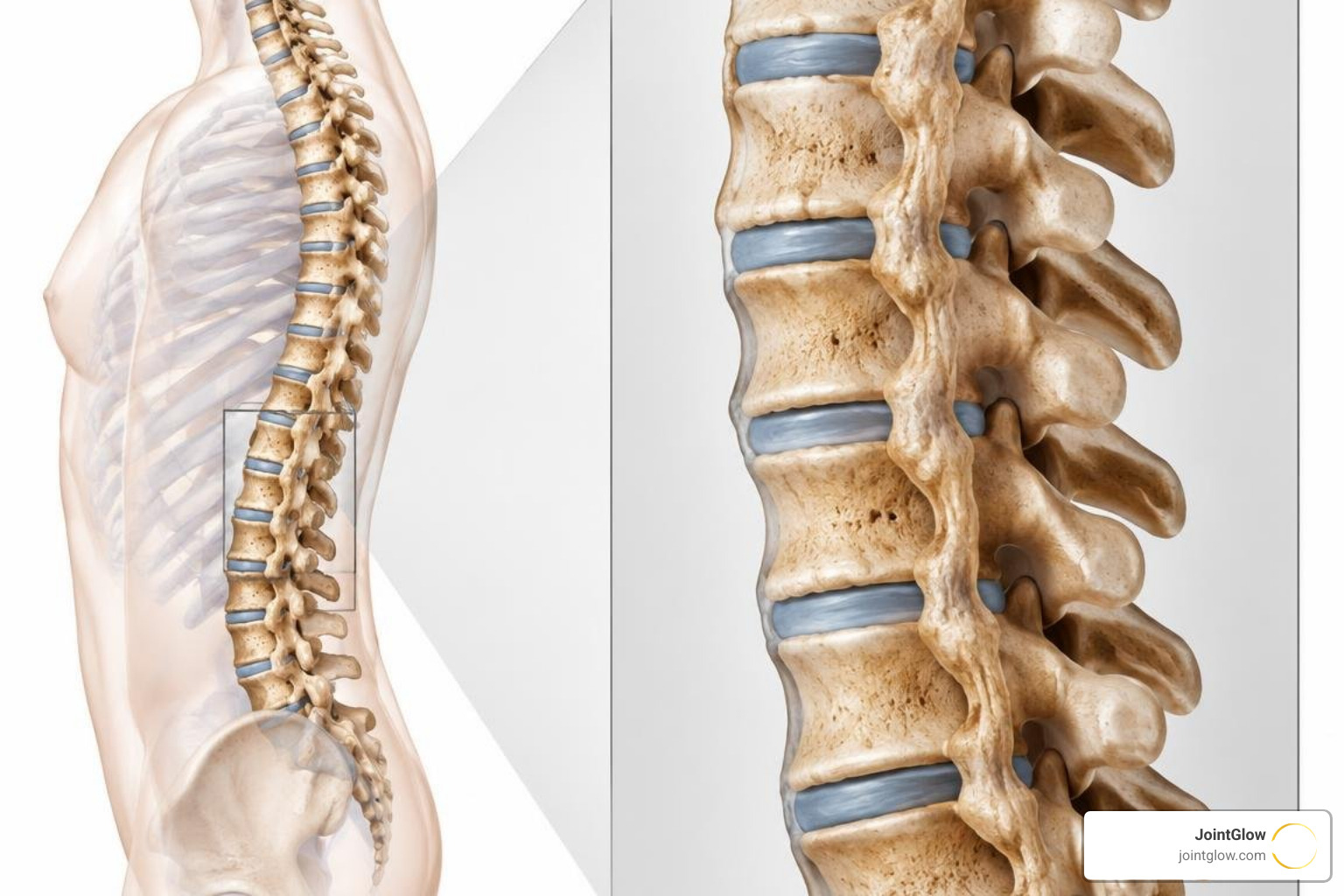
The central structural mystery of ankylosing spondylitis is its ability to cause both bone loss (osteoporosis) and excessive, pathological new bone formation at the exact same time. This destructive process centers on the enthesis—the specialized anatomical zone where tendons, ligaments, and joint capsules insert directly into bone.
Spinal entheses are exposed to constant mechanical loading. In someone predisposed to AS, normal mechanical micro-damage triggers an excessive inflammatory response rather than clean, quiet tissue repair. Research decoding the inflammatory-osteogenic axis in AS shows that active inflammation primes local mesenchymal stem cells to begin forming abnormal structural bone bridges.
How Ankylosing Spondylitis Inflammation Causes Bone Fusion
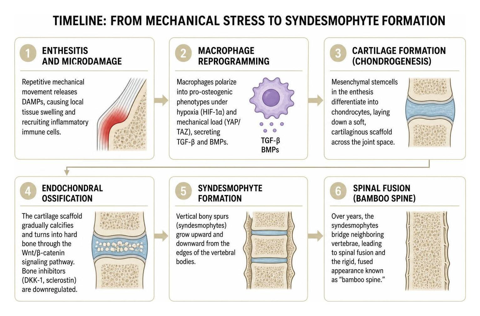
The path from localized joint pain to spinal rigidity involves a distinct cascade of biological events:
- Enthesitis and Microdamage: Repetitive mechanical movement releases damage-associated molecular patterns (DAMPs), causing local tissue swelling and recruiting inflammatory immune cells.
- Macrophage Reprogramming: As detailed in macrophage-mediated TGF-beta and BMP signaling research, local macrophages undergo polarization into hybrid pro-osteogenic phenotypes. Under the influence of local hypoxia (HIF-1α) and mechanical load (YAP/TAZ pathways), they secrete transforming growth factor-beta (TGF-β) and bone morphogenetic proteins (BMPs).
- Cartilage Formation (Chondrogenesis): Mesenchymal stem cells in the enthesis differentiate into chondrocytes, laying down a soft, cartilaginous scaffold across the joint space.
- Endochondral Ossification: This cartilage scaffold gradually calcifies and turns into hard bone through the Wnt/β-catenin signaling pathway. Natural bone inhibitors such as Dickkopf-1 (DKK-1) and sclerostin are locally downregulated.
- Syndesmophyte Formation: These vertical bony spurs (syndesmophytes) slowly grow upward and downward from the edges of the vertebral bodies. Over years, they bridge neighboring vertebrae, ultimately producing the rigid, fused appearance classically referred to on X-rays as “bamboo spine.”
Systemic Complications Beyond the Spine: Eyes, Heart, and Lungs
Because the underlying immune drivers circulate through the entire bloodstream, ankylosing spondylitis can trigger complications far outside the spine and pelvis:
- Acute Anterior Uveitis (Iritis): Occurring in roughly 25% to 40% of patients, this sudden eye inflammation causes severe pain, redness, light sensitivity (photophobia), and blurred vision. It requires urgent medical care to avoid permanent vision changes.
- Cardiovascular Complications: Chronic systemic inflammation can affect the root of the aorta, leading to aortitis, aortic valve regurgitation, and cardiac electrical conduction abnormalities such as heart block.
- Chest Wall Restriction and Pulmonary Issues: Enthesitis where the ribs meet the spine (costovertebral joints) can make deep breathing painful, causing reduced chest expansion. Over decades, rare apical lung fibrosis can develop.
- Gastrointestinal Inflammation: Up to 10% of patients with AS develop clinically recognized inflammatory bowel disease (Crohn’s disease or ulcerative colitis), while over 50% demonstrate microscopic, subclinical intestinal inflammation.
- Spinal Fractures: Paradoxically, while new bone fuses the outside of the spine, the interior bone often becomes osteoporotic. Fused, brittle spines act like long, rigid bones that can fracture easily even after minor falls.
Recognizing Symptoms and Clinical Diagnosis
Catching spinal arthritis early is vital for preserving long-term posture and spinal mobility. Unfortunately, because low back pain is so common in the general population, the average diagnostic delay for ankylosing spondylitis historically ranged between five and eight years. Knowing what makes inflammatory back pain unique is the key to prompt medical evaluation.
When assessing individuals with persistent musculoskeletal issues, clinicians must differentiate between mechanical strain and systemic inflammatory disease. You can review how we approach related joint conditions and chronic inflammatory disorders to understand these clinical distinctions.
| Feature | Inflammatory Back Pain (AS) | Mechanical Back Pain (Strain/Disc) |
|---|---|---|
| Age of Onset | Typically < 40–45 years old | Any age (often increases with age) |
| Onset Style | Gradual and insidious (over months) | Sudden or following a specific physical injury |
| Morning Stiffness | Lasts > 30–60 minutes, severe | Short-lived (< 30 minutes), mild |
| Effect of Exercise | Improves pain and stiffness | Worsens pain; relieved by rest |
| Nocturnal Pain | Common, wakes patient in second half of night | Rare, usually relieved by lying down |
| Buttock Pain | Alternating, deep buttock pain (sacroiliitis) | Unilateral, shooting down one leg (radiculopathy) |
Early Warning Signs vs Mechanical Back Pain
The classic hallmark of ankylosing spondylitis is back pain that defies ordinary logic: resting makes it worse, while moving around makes it better. People often describe waking up at 3:00 or 4:00 AM needing to stretch, walk around the bedroom, or take a hot shower just to loosen up their lower back.
Stiffness typically lasts well over 30 to 60 minutes after getting out of bed. Another early warning sign is alternating buttock pain, caused by inflammation in the bilateral sacroiliac (SI) joints—the weight-bearing connection between the pelvis and the base of the spine. Heel pain (plantar fasciitis or Achilles tendinitis) and unprovoked swelling in large joints like the hips or knees are also frequent early clues.
Diagnostic Workup: Imaging, Blood Tests, and Biomarkers
A complete medical evaluation combines specialized physical exams, blood work, and imaging studies:
- Sacroiliac Joint and Spine Imaging: Conventional X-rays can reveal established sacroiliitis, sclerosis, and syndesmophytes. However, early disease may not show up on plain films for several years. Magnetic Resonance Imaging (MRI) is the gold standard for early detection because it highlights active bone marrow edema and enthesitis long before permanent structural bone changes occur.
- Blood Biomarkers: Inflammatory markers such as Erythrocyte Sedimentation Rate (ESR) and C-Reactive Protein (CRP) are elevated in roughly 60% to 70% of active cases. Clinicians also check for matrix metalloproteinase-3 (MMP-3), a marker of ongoing joint and cartilage turnover.
- Genetic Testing: Testing for the HLA-B27 genetic marker provides strong supportive diagnostic data when paired with clinical symptoms.
- Clinical Disease Activity Scoring: Tools such as the Bath Ankylosing Spondylitis Disease Activity Index (BASDAI) and the Ankylosing Spondylitis Disease Activity Score (ASDAS) allow physicians to quantitatively measure pain, fatigue, and joint swelling over time.
For an extensive review of modern diagnostic strategies, clinical papers and updates on disease biomarkers and diagnostic imaging offer detailed breakdowns of these criteria.
Comprehensive Management Strategies and Emerging Therapies
Managing ankylosing spondylitis requires a proactive, multidisciplinary approach. The primary goals are simple: extinguish active inflammation, relieve chronic pain, preserve physical posture, and prevent irreversible spinal fusion.
Medical Management and Non-Invasive LDRT
Traditional medical care follows a stepwise approach tailored to disease severity:
- Nonsteroidal Anti-Inflammatory Drugs (NSAIDs): Medications such as naproxen, celecoxib, and indomethacin serve as the first-line pharmacologic baseline, dampening prostaglandin synthesis and easing daily morning stiffness.
- Biologic DMARDs: For moderate-to-severe disease that does not fully respond to NSAIDs, biologic agents targeting Tumor Necrosis Factor (TNF-α blockers such as infliximab, adalimumab, etanercept) or Interleukin-17 (IL-17A inhibitors such as secukinumab and ixekizumab) are prescribed to calm systemic inflammation.
- Targeted Small Molecules: Oral Janus kinase (JAK) inhibitors (such as tofacitinib and upadacitinib) block intracellular cytokine signaling downstream.
- Structured Physical Therapy and Daily Posture Training: Dedicated stretching routines, swimming, and back-strengthening exercises are non-negotiable parts of living with AS, helping preserve thoracic expansion and spinal mobility.
In addition to traditional medical therapies, specialized non-pharmacologic interventions play an important role in controlling localized, refractory joint inflammation. At JointGlow, we offer non-invasive low-dose radiation therapy (LDRT) as an effective, Medicare Part B (Excludes Medicare Advantage) covered option for individuals dealing with chronic musculoskeletal inflammation. LDRT delivers ultra-low, therapeutic doses of ionizing energy directly to inflamed joint spaces.
Rather than destroying tissue like high-dose oncologic radiation, low-dose radiation operates on a cellular level: it dampens pro-inflammatory macrophage activity, lowers endothelial adhesion molecules, and stimulates local anti-inflammatory cytokines (such as TGF-β1 and IL-10). This process helps biologically reset the local joint environment, delivering durable pain relief without the systemic side effects, injections, or surgical downtime often seen with traditional approaches.
Novel Research Frontiers: AIM2 Inhibitors and Immunomodulation
The future of spondyloarthritis care is advancing rapidly beyond standard cytokine suppression. Because traditional biologics frequently control pain without completely stopping progressive syndesmophyte growth, current research is zeroing in on upstream and bone-specific cellular targets:
- Targeted Mitochondrial Antioxidants (MitoQ): Emerging studies show that scavenging mitochondrial reactive oxygen species prevents the release of cytosolic mtDNA, blocking AIM2 inflammasome activation before inflammatory cascades even start.
- Smooth-Agonist/Antagonist (SMO) Pathways: By disrupting the hedgehog-SMO signaling axis in local entheseal stem cells, researchers aim to halt abnormal chondrocyte differentiation and stop new bone bridges from forming.
- Macrophage Reprogramming Therapies: Novel small molecules designed to block YAP/TAZ mechanotransduction and inhibit latent TGF-β activation are in development to decouple joint inflammation from pathological ossification.
You can stay informed on these cutting-edge therapeutic developments by exploring our ongoing clinical research on advanced arthritis interventions.
Frequently Asked Questions About Ankylosing Spondylitis
When should I see a rheumatologist for persistent lower back pain?
You should consult a rheumatologist if your low back or buttock pain has lasted for more than three months, began before the age of 45, is worst in the morning (lasting over 30 to 60 minutes), and noticeably improves with physical activity rather than rest. Urgent medical evaluation is essential if you experience red-flag symptoms such as sudden eye redness and pain (possible uveitis), unexplained weight loss, chronic gastrointestinal distress, or if you have a first-degree relative diagnosed with ankylosing spondylitis or related autoimmune conditions.
Can ankylosing spondylitis be cured or completely stopped?
While ankylosing spondylitis does not currently have a permanent medical cure, it is highly manageable. Modern treatment strategies—combining early biologic DMARDs, targeted anti-inflammatory interventions, dedicated physical therapy, and healthy lifestyle choices—allow many patients to achieve long-term clinical remission. Early, proactive treatment dramatically lowers disease activity, prevents permanent postural changes, and protects long-term quality of life.
Does testing positive for HLA-B27 mean I will develop ankylosing spondylitis?
No, carrying the HLA-B27 gene does not mean you are guaranteed to develop the disease. While over 85% to 90% of white individuals with ankylosing spondylitis carry the HLA-B27 marker, only about 5% of all people who test positive for HLA-B27 will ever develop AS. The gene represents a genetic vulnerability that requires additional environmental triggers—such as gut dysbiosis, biomechanical entheseal stress, or immune dysregulation—to spark active chronic inflammation.
Conclusion
Living with ankylosing spondylitis can feel challenging, but understanding the underlying mechanisms of chronic inflammation puts you firmly in control of your health journey. From genetic predispositions like HLA-B27 and cellular cascades such as the AIM2 inflammasome to the biomechanics of enthesitis, each piece of the scientific puzzle helps us design better, more targeted ways to protect your mobility.
Achieving lasting relief requires an active, multidisciplinary partnership combining accurate diagnosis, smart medical therapies, dedicated daily movement, and advanced anti-inflammatory treatments. At JointGlow, our mission is to support your journey toward lasting comfort through safe, non-invasive therapies. To learn more about what to anticipate on your treatment journey, explore our detailed timeline for achieving maximum pain relief.
