When Sudden Hip Pain Makes Walking Impossible, Stop and Get Help
If sudden hip pain can’t walk describes what is happening now, do not try to push through it. Stop putting weight on the leg, sit or lie down somewhere safe, and ask someone to help.
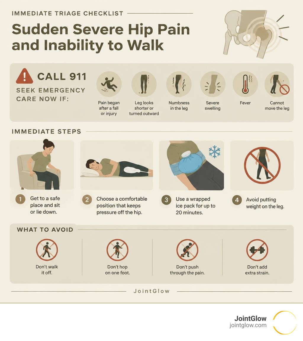
Call 911 or seek emergency care now if the pain began after a fall or injury, your leg looks shorter or turned outward, you have numbness, severe swelling, fever, or cannot move the leg. These signs can point to a fracture, dislocation, infection, or another urgent problem.
For pain without obvious injury, avoid “walking it off.” Use a wrapped ice pack for up to 20 minutes, protect the hip from further strain, and arrange prompt medical assessment, especially if you cannot bear weight. Sudden immobility can come from the hip joint itself, but it can also be referred from the spine, pelvis, or nearby nerves.
I am Dr. Koneru, and the next steps will help you separate a possible emergency from a problem that needs urgent but non-emergency care.
Immediate Steps: What to Do (and What to Avoid) Right Now
When sharp, immobilizing pain hits your hip, your body is sending an unmistakable signal to offload the joint immediately. The very first priority is fall prevention. If you are standing, safely lower yourself to the nearest stable surface—a sturdy chair, a sofa, or even the floor if no seating is nearby. Do not attempt to hop on one foot across the room, as this drastically increases the risk of a secondary fall and further orthopedic trauma.
Once seated or lying down, adopt a position that minimizes mechanical pressure on the acetabulofemoral joint. Most individuals find the greatest relief lying on their non-injured side with pillows placed securely between their knees and ankles to keep the pelvis neutral. Alternatively, lying on your back with a rolled towel or pillow placed under your knees reduces tension on the iliopsoas muscle and the anterior joint capsule.

Stop Moving: Why You Should Never Try to Walk It Off
The old adage of “walking it off” is dangerous when it comes to acute joint failure. Forcing weight onto a compromised hip can turn an incomplete stress fracture into a displaced femoral neck fracture, permanently altering joint geometry and jeopardizing blood supply to the femoral head.
If the pain is caused by an acute labral tear, continuing to step on the leg can grind torn fibrocartilage between the femoral head and acetabulum, causing rapid secondary damage to the articular surface. Following clinical guidance on avoiding joint strain is critical: offload the leg completely until you have a clear medical assessment.
First-Aid Protocol: RICE and Pain Management
For non-emergency acute flare-ups and suspected soft-tissue injuries, applying a structured first-aid protocol helps control localized swelling and down-regulate pain receptors:
- Rest: Discontinue all physical activity and avoid bearing weight on the affected limb.
- Ice: Apply an ice pack wrapped in a thin towel to the painful area for 15 to 20 minutes every 2 to 3 hours. Never apply bare ice directly to the skin.
- Compression: If the pain is localized to the groin or outer thigh, wearing supportive compression shorts can help stabilize the surrounding musculature, though you should avoid tight elastic wraps that restrict arterial blood flow.
- Elevation: When lying down, elevate the leg slightly using pillows to encourage venous return and decrease localized edema.
For medical pain relief, follow clinician guidance regarding over-the-counter anti-inflammatories (such as ibuprofen or naproxen) or paracetamol. Avoid masking severe pain with heavy analgesics simply to force yourself to walk.
Emergency Signs: When Sudden Hip Pain Can’t Walk Requires 911 Care
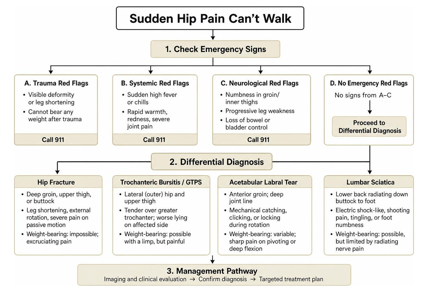
Certain clinical presentations demand immediate emergency medical intervention. Reviewing established guidance on emergency hip pain symptoms reveals several clear red flags:
- Visible Deformity or Leg Shortening: If your affected leg appears visibly shorter than the other or is rotated abnormally outward, this strongly indicates a displaced hip fracture or acute dislocation.
- Inability to Bear Any Weight After Trauma: Following a fall or high-impact accident, the complete inability to stand warrants immediate evaluation according to NHS protocols on broken hip trauma. Do not attempt to drive yourself; call for emergency medical transport.
- Systemic Red Flags (Septic Arthritis): Severe joint pain accompanied by a sudden high fever, chills, rapid localized warmth, or redness points to an intra-articular infection, which can destroy joint cartilage within hours if untreated.
- Neurological Deficits: Sudden numbness in the groin or inner thighs (saddle anesthesia), progressive leg weakness, or loss of bowel or bladder control are signs of cauda equina syndrome and require emergency spinal decompression surgery.
Sudden Hip Pain Can’t Walk: Key Causes and Differential Diagnosis
To understand why your hip suddenly stopped supporting your weight, we must differentiate between structural bone disruption, soft-tissue inflammation, and neuropathic referral patterns.
| Condition | Primary Pain Location | Key Identifying Symptoms | Weight-Bearing Capacity |
|---|---|---|---|
| Hip Fracture | Deep groin, upper thigh, or buttock | Leg shortening, external rotation, severe pain on passive motion | Completely impossible; excruciating pain |
| Trochanteric Bursitis / GTPS | Lateral (outer) hip and upper thigh | Tenderness over greater trochanter; worse lying on affected side | Possible with a limp, but sharply painful |
| Acetabular Labral Tear | Anterior groin; deep joint line | Mechanical catching, clicking, or locking sensations during rotation | Variable; sharp pain on pivoting or deep flexion |
| Lumbar Sciatica | Lower back radiating down buttock to foot | Electric shock-like, shooting pain, tingling, or foot numbness | Possible, but limited by radiating nerve pain |
Acute Fractures, Labral Tears, and Avascular Necrosis
Structural bone and cartilage failures represent the most severe intra-articular causes of sudden immobility. A femoral neck or intertrochanteric fracture can happen not only after major trauma, but also spontaneously in older adults with underlying osteoporosis.
Acetabular labral tears occur when the ring of cartilage securing the ball of the hip joint is torn due to sudden twisting, hyperflexion, or underlying femoroacetabular impingement (FAI). This often manifests as an abrupt, sharp groin pain accompanied by a mechanical catching sensation.
Avascular necrosis (osteonecrosis) occurs when the local blood supply to the femoral head is compromised, leading to cellular bone death and microfractures that eventually cause the articular surface to collapse. For a deeper look at these conditions, consult this expert overview of structural hip injuries.
Severe Inflammation: Greater Trochanteric Pain Syndrome and Tendonitis
Not all sudden hip pain originates inside the joint socket. Greater Trochanteric Pain Syndrome (GTPS) is a broad clinical diagnosis that includes trochanteric bursitis and gluteal tendinopathy. Interestingly, while outer hip pain is frequently diagnosed as bursitis, clinical studies indicate that over 90% of outer hip pain is actually driven by tendinopathy of the gluteus medius or minimus, or tight myofascial structures like the iliotibial band, rather than isolated fluid inflammation of the bursa.
Acute flare-ups of iliopsoas tendonitis or adductor strains can also cause sudden, sharp anterior hip pain when lifting the knee. You can review detailed information on various underlying hip conditions to better identify these soft-tissue patterns.
Referred Pain: Sciatica, Hernias, and Pelvic Conditions
Because the hip receives sensory innervation from lumbar nerve roots (L1 through S1), pathology outside the joint frequently mimics acute hip injuries.
- Lumbar Radiculopathy (Sciatica): A herniated lumbar disc compressing the L4 or L5 nerve root creates radiating, shooting pain through the buttock and hip down the leg.
- Piriformis Syndrome: Spasm of the deep piriformis muscle compresses the sciatic nerve, triggering acute hip and gluteal immobility.
- Inguinal Hernias: A protrusion of abdominal tissue through the inguinal canal produces sharp, sudden groin and anterior hip pain exacerbated by coughing or standing.
- Gynecological and Pelvic Issues: Endometriosis, ovarian cysts, and pelvic inflammatory disease (PID) can refer severe pain directly to the anterior pelvis and deep hip joint.
Diagnostic Tests and Medical Evaluation
Arriving at an accurate diagnosis requires a structured orthopedic workup that moves systematically from physical examination to advanced imaging.
Clinical Examination and Functional Mobility Assessments
During your clinical evaluation, the physician will perform targeted orthopedic tests to localize the anatomical source of pain:
- Stinchfield Test: The patient performs an active straight-leg raise against downward resistance. Pain in the groin confirms intra-articular hip joint pathology.
- FABER (Flexion, Abduction, External Rotation) Test: Also known as Patrick’s test, this assesses sacroiliac joint pathology and anterior hip capsule irritation.
- FADIR (Flexion, Adduction, Internal Rotation) Test: This test checks for femoroacetabular impingement (FAI) and acetabular labral tears by compressing the femoral neck against the acetabular rim.
- Neurovascular Exam: Testing peripheral pulses, sensation across dermatomes, and deep tendon reflexes rules out vascular compromise and acute nerve root compression.
Advanced Diagnostic Imaging: X-Rays, MRI, and Ultrasound
Imaging provides objective confirmation of underlying bone and soft-tissue architecture:
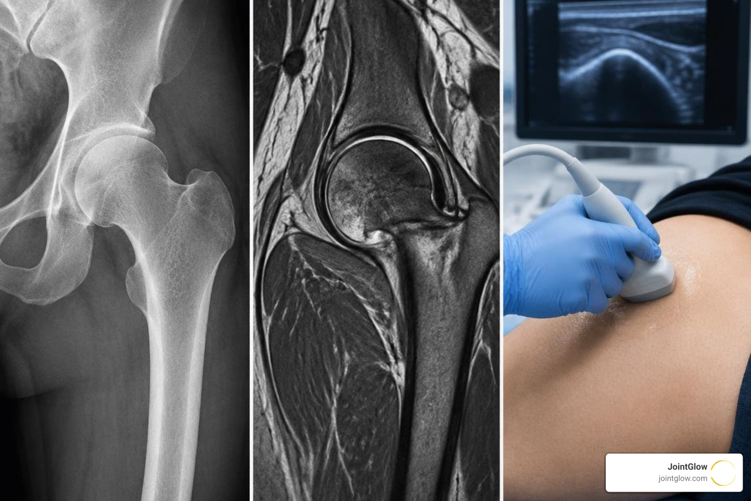
- Plain Radiography (X-rays): The standard initial test, capturing AP pelvis and frog-leg lateral views to identify acute fractures, joint space narrowing, and bone osteophytes.
- Magnetic Resonance Imaging (MRI / MR Arthrogram): The gold standard for detecting occult stress fractures, avascular necrosis, bone marrow edema, and acetabular labral tears.
- Diagnostic Musculoskeletal Ultrasound: Excellent for visualizing soft-tissue fluid collections, trochanteric bursitis, and dynamic tendon movement in real time.
- Diagnostic Joint Aspiration: If septic arthritis or crystalline arthropathy (gout) is suspected, synovial fluid is aspirated and sent for cell count, Gram stain, and culture.
JointGlow Treatment Considerations for Chronic Joint Inflammation
While acute surgical emergencies such as displaced fractures or septic joints require immediate operating room intervention, many cases of sudden immobility stem from acute exacerbations of underlying chronic degenerative and inflammatory joint disease.
Non-Invasive Low-Dose Radiotherapy for Chronic Joint Inflammation
When chronic hip inflammation from conditions like osteoarthritis or persistent greater trochanteric pain syndrome causes severe flares that impair everyday mobility, advanced non-invasive options offer durable relief.
At JointGlow, we provide specialized low-dose radiotherapy (LDRT), a clinically proven, non-invasive treatment covered by Medicare. LDRT works by delivering ultra-low doses of ionizing radiation directly to the inflamed joint tissues. Rather than destroying tissue (as in oncological radiation), LDRT modulates local inflammatory pathways:
- It induces apoptosis in pro-inflammatory immune cells (macrophages and activated lymphocytes).
- It down-regulates inflammatory cytokines like IL-1, IL-6, and TNF-alpha.
- It resets the microenvironment of the joint, facilitating drug-free, non-surgical symptom relief without downtime.
Understanding how radiotherapy resets joint inflammation allows patients dealing with recurrent, debilitating flare-ups to achieve lasting mobility without the systemic side effects of chronic NSAIDs or the risks of repeated intra-articular steroid injections.
When Specialist Intervention Is Needed
When conservative modalities and advanced anti-inflammatory therapies are insufficient, orthopedic surgical interventions may be necessary:
- Hip Arthroscopy: A minimally invasive procedure used to repair torn labral tissue and shave down impinging bone spurs in FAI.
- Open Reduction and Internal Fixation (ORIF): Surgical stabilization of hip fractures using specialized cannulated screws, intramedullary nails, or dynamic hip plates.
- Total Hip Arthroplasty (Hip Replacement): Indicated for end-stage joint destruction where bone-on-bone osteoarthritis or advanced avascular necrosis eliminates natural joint function.
Recovery Timeline: What to Expect When Sudden Hip Pain Can’t Walk Stops You
Recovery duration depends heavily on the underlying diagnosis and the severity of tissue damage:
- Grade I Muscle/Flexor Strain: Mild micro-tearing typically resolves within 1 to 3 weeks with conservative RICE and progressive stretching.
- Grade II Moderate Strain: Partial muscle tears generally require 4 to 8 weeks of dedicated physical rehabilitation.
- Grade III Complete Tear or Labral Surgery: Requires extensive medical intervention, with rehabilitation protocols lasting 3 to 6 months.
- Hip Fracture Repair: Bone union typically requires 8 to 12 weeks, with functional gait recovery and strength rebuilding continuing for 6 to 12 months.
Frequently Asked Questions About Sudden Severe Hip Pain
Is sudden hip pain that prevents walking always an emergency?
Not every case requires an ambulance, but any sudden inability to bear weight warrants urgent medical evaluation within 24 hours. If the pain follows a fall, features visible leg deformity, or is accompanied by fever, chills, or sudden leg numbness, it is a true medical emergency that requires immediate 911 or emergency department care.
Can a pinched nerve in the back cause sudden, severe hip pain?
Yes. Compression of the L4, L5, or S1 nerve roots in the lumbar spine (commonly caused by acute disc herniations or spinal stenosis) frequently radiates severe, sharp, or burning pain directly into the buttock, outer hip, and groin, mimicking an acute hip injury.
How can I safely move to a doctor or hospital if I can’t put weight on my leg?
If a fracture or serious traumatic injury is suspected, do not attempt to transport yourself; call emergency services (911) for professional paramedic assistance. If non-emergency transport is appropriate, use a sturdy pair of crutches or a walker to offload the affected leg completely, or have two assistants support your weight without putting your injured foot on the ground.
Conclusion
Experiencing sudden, severe hip pain that makes walking impossible is a clear signal that your body requires immediate rest and professional evaluation. By quickly recognizing emergency red flags—such as trauma-induced deformity, fever, or neurological deficits—you can take the right actions to protect your joint from secondary damage.
For chronic, recurrent inflammatory conditions and severe degenerative joint pain that disrupt your mobility, explore advanced non-invasive solutions at JointGlow to discover how targeted therapies can restore your comfort, function, and independence.
